. 2012 Jan;32(1):57-69.
doi: 10.1038/jcbfm.2011.106. Epub 2011 Jul 27.

Tissue-type plasminogen activator has a neuroprotective effect in the ischemic brain mediated by neuronal TNF-α

Affiliations

Tissue-type plasminogen activator has a neuroprotective effect in the ischemic brain mediated by neuronal TNF-α

Woldeab B Haile et al. J Cereb Blood Flow Metab. 2012 Jan.

Abstract

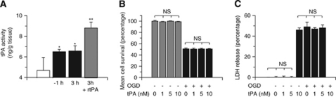
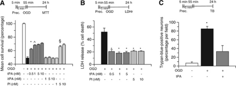
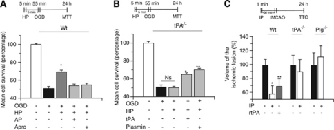
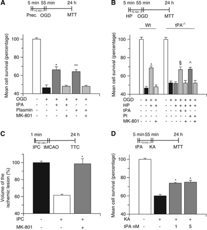
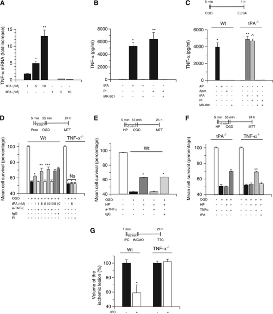
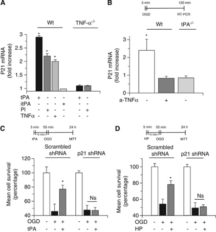
Cerebral cortical neurons have a heightened sensitivity to hypoxia and their survival depends on their ability to accommodate to changes in the concentration of oxygen in their environment. Tissue-type plasminogen activator (tPA) is a serine proteinase that activates the zymogen plasminogen into plasmin. Hypoxia induces the release of tPA from cerebral cortical neurons, and it has been proposed that tPA mediates hypoxic and ischemic neuronal death. Here, we show that tPA is devoid of neurotoxic effects and instead is an endogenous neuroprotectant that renders neurons resistant to the effects of lethal hypoxia and ischemia. We present in vitro and in vivo evidence indicating that endogenous tPA and recombinant tPA induce the expression of neuronal tumor necrosis factor-α. This effect, mediated by plasmin and the N-methyl-D-aspartate receptor, leads to increased expression of the cyclin-dependent kinase inhibitor p21 and p21-mediated development of early hypoxic and ischemic tolerance.


Figures

Figure 1
Figure 1
Tissue-type plasminogen activator (tPA) is not neurotoxic to cerebral cortical neurons. (A) Mean concentration of active tPA in the brain of wild-type (Wt) mice either under non-ischemic conditions (white bar) or 1 or 3 hours after transient occlusion of the middle cerebral artery (tMCAO; black bars), or 3 hours after tMCAO and the intravenous administration of recombinant tPA (rtPA; gray bar); n=8 for each group. Data represent mean±s.d. *P<0.05 versus non-ischemic brains. **P<0.01 versus untreated ischemic brains 1 and 3 hours after tMCAO. Mean neuronal survival determined by the 3-(4,5-dimethylthiazol-2-yl)-2,5-diphenyltetrazolium bromide assay (B) and quantification of lactate dehydrogenase (LDH) release into the media (C) in Wt cerebral cortical neurons incubated with tPA 0 to 10 nmol/L and either maintained under normoxia (gray bars) or simultaneously exposed to oxygen–glucose deprivation (OGD) conditions for 55 minutes (black bars); n=15 for each group. Data represent mean±s.d. NS, nonsignificant.
Figure 2
Figure 2
Proteolytically active tissue-type plasminogen activator (tPA) induces hypoxic tolerance. Mean neuronal survival determined by the 3-(4,5-dimethylthiazol-2-yl)-2,5-diphenyltetrazolium bromide (MTT) assay (A) and quantification of lactate dehydrogenase (LDH) release into the media (B) in wild-type (Wt) cerebral cortical neurons incubated during 5 minutes with 0 to 10 nmol/L of either active tPA or proteolytically inactive tPA (itPA), or plasmin (Pl), followed 5 minutes later by exposure to 55 minutes of oxygen–glucose deprivation (OGD) conditions; n=12 for each group. Data represent mean±s.d. (A) *P<0.01 versus cells maintained under OGD conditions without preconditioning with tPA, ^P<0.05 versus cells treated with tPA 0.5 nmol/L before exposure to lethal OGD conditions, and §P<0.01 versus untreated neurons exposed to OGD conditions. (B) *P<0.01 versus cells maintained under OGD conditions without previous incubation with tPA or Pl. Prec. denotes preconditioning and indicates the moment when neurons were treated. (C) Percentage of trypan blue-positive cells/field in Wt neuronal cultures maintained under normoxia (white bars) or exposed to lethal OGD conditions for 55 minutes (black bars) or incubated with tPA 5 nmol/L 5 minutes before exposure to 55 minutes of OGD conditions (gray bars); n=10 for each group. Data represent mean±s.d. *P<0.01 versus cells maintained under OGD conditions without treatment. Prec. denotes preconditioning and indicates the time of treatment with tPA. LDHr, LDH release assay; TB, trypan blue exclusion assay.
Figure 3
Figure 3
Endogenous tissue-type plasminogen activator (tPA) induces hypoxic tolerance. Mean cell survival determined by the 3-(4,5-dimethylthiazol-2-yl)-2,5-diphenyltetrazolium bromide (MTT) assay in wild-type (Wt; A) and tPA-deficient (tPA−/−; B) neurons exposed to 5 minutes of oxygen–glucose deprivation (OGD) conditions (hypoxic preconditioning, HP), followed 5 minutes later by 55 minutes of OGD. A subgroup of neurons was incubated during the preconditioning phase with either α2-antiplasmin 100 nmol/L (AP) or aprotinin 1.4 μmol/L (Apro), or tPA 5 nmol/L or plasmin 10 nmol/L. HP denotes the moment when cells were exposed to sublethal hypoxia; n=12 for each observation. Data represent mean cell survival±s.d. (A) *P<0.01 versus cells exposed to OGD without hypoxic preconditioning. (B) *P<0.01 versus tPA−/− neurons preconditioned in the absence of tPA and **P<0.01 versus tPA−/− neurons preconditioned in the absence of plasmin. (C) Mean percentage decrease in the volume of the ischemic lesion compared with non-preconditioned mice (black bars) in Wt, tPA−/−, and plasminogen (Plg−/−) mice preconditioned by exposure to 1 minute of low oxygen concentration (ischemic preconditioning, IP), followed 60 minutes later by transient middle cerebral artery occlusion (tMCAO). Black bars depict mice undergoing tMCAO without preconditioning. A subgroup of Wt mice was preconditioned with recombinant tPA (rtPA) intravenously administered 60 minutes before tMCAO (gray bar); n=12 for each observation. Data represent mean percentage compared with stroke volume in non-preconditioned brains±s.d. *P<0.01 compared with non-preconditioned Wt mice. **P<0.05 versus non-preconditioned Wt mice. Ns, non significant; TTC, triphenyltetrazolium chloride staining.
Figure 4
Figure 4
The N-methyl--aspartate receptor (NMDAR) is required for the neuroprotective effect of tissue-type plasminogen activator (tPA). (A) Mean cell survival in wild-type (Wt) neurons treated with tPA 5 nmol/L or plasmin 10 nmol/L, either alone or in combination with the NMDAR antagonist MK-801 10 μ, followed 5 minutes later by exposure to 55 minutes of oxygen–glucose deprivation (OGD) conditions; n=12 for each observation. *P<0.01 versus cells cotreated with tPA and MK-801. **P<0.01 versus cells cotreated with plasmin and MK-801. (B) Mean cell survival in Wt and tPA−/− neurons exposed to 5 minutes of OGD (hypoxic preconditioning, HP), followed 5 minutes later by incubation under OGD conditions for 55 minutes. A subgroup of tPA−/− cells was treated with tPA 5 nmol/L or plasmin 10 nmol/L either alone or in combination with MK-801 10 μmol/L; n=10 for each observation. Data represent mean±s.d. *P<0.01 versus cells cotreated with MK-801. §P<0.01 versus tPA−/− neurons preconditioned either in absence of tPA or with a combination of tPA and MK-801. ^P<0.01 versus preconditioned and non-preconditioned tPA−/− neurons and versus tPA−/− neurons cotreated with plasmin and MK-801 during the preconditioning phase. HP denotes the moment when cells were treated with tPA or plasmin alone or in combination with MK-801. Indicates the moment when cells were exposed to sublethal hypoxia. (C) Mean percentage decrease in the volume of the ischemic lesion in Wt mice either left untreated or injected with MK-801 1 μg/kg intraperitoneally, followed by ischemic preconditioning and transient occlusion of the middle cerebral artery (tMCAO) 1 hour later; n=10; *P<0.01 versus mice preconditioned without MK-801. (D) Mean cell survival in Wt neurons incubated with tPA 1 or 5 nmol/L, followed 5 minutes later by exposure to kainic acid (KA) 250 μmol/L; n=10; P<0.01 versus neurons exposed to KA without preconditioning with tPA. IPC, ischemic preconditioning.
Figure 5
Figure 5
Tumor necrosis factor (TNF)-α mediates the neuroprotective effect of tissue-type plasminogen activator (tPA). (A) TNF-α mRNA expression in wild-type (Wt) neurons incubated with active tPA or inactive tPA (itPA) 1 to 10 nmol/L; n=12 for each observation. Data represent mean fold increase in TNF-α mRNA compared with control cells±s.d. *P<0.01 versus neurons treated with tPA 1 nmol/L. **P<0.01 versus neurons treated with tPA 5 nmol/L. (B) TNF-α concentration (pg/ml) in the culture media of Wt neurons incubated with tPA 5 nmol/L or plasmin (Pl) 10 nmol/L either alone or in combination with MK-801 10 μmol/L; n=12 for each observation. *P<0.01 versus neurons either left untreated or cotreated with tPA and MK-801. **P<0.01 versus neurons either left untreated or cotreated with Pl and MK-801. (C) Mean TNF-α concentration (pg/ml) in the media of Wt and tPA−/− neurons exposed to 5 minutes of oxygen–glucose deprivation (OGD) conditions (hypoxic preconditioning) alone or in the presence of either α2-antiplasmin 100 nmol/L (AP) or aprotinin 1.4 μmol/L (Apro) or tPA 5 nmol/L or Pl 10 nmol/L or MK-801 10 μmol/L; n=12 for each observation. Lines denote s.d. *P<0.01 versus neurons treated with either AP or Apro or MK-801. ** and ^P<0.01 versus untreated tPA−/− neurons. (D) Mean cell survival in Wt and TNF-α−/− neurons incubated with tPA 1 to 10 nmol/L, or with a combination of tPA 1 to 10 nmol/L and either anti-TNF-α-blocking antibodies 0.4 μg/ml (a-TNF-α) or an isotype immunoglobulin G (IgG) control (dark gray bars), or with Pl alone 5 nmol/L, followed 5 minutes later by exposure to lethal OGD conditions; n=12 for each observation. Lines denote s.d. *P<0.01 versus neurons cotreated with tPA 1 nmol/L and a-TNF-α antibodies. **P<0.01 versus neurons cotreated with tPA 5 nmol/L and a-TNF-α antibodies. ***P<0.01 versus neurons cotreated with tPA 10 nmol/L and a-TNF-α antibodies. Prec. indicates preconditioning and denotes the moment when cells where treated. (E) Mean cell survival in Wt neurons exposed to 5 minutes of OGD (hypoxic preconditioning, HP) alone or in the presence of either anti-TNF-α-neutralizing antibodies or an IgG isotype control, followed 5 minutes later by exposure to lethal OGD conditions; n=12 for each observation. *P<0.01 versus neurons treated with a-TNF-α antibodies during the preconditioning phase. Lines denote mean±s.d. (F) Mean cell survival in tPA−/− and TNF-α−/− neurons exposed to 5 minutes of OGD conditions (HP) either alone or in combination with tPA 5 nmol/L or TNF-α 20 ng/ml, followed 5 minutes later by exposure to 55 minutes OGD; n=10 for each observation. *P<0.01 versus preconditioned and non-preconditioned tPA−/− neurons. **P<0.01 versus non-preconditioned and preconditioned TNF-α−/− neurons and versus TNF-α−/− neurons treated with tPA during the preconditioning phase. Lines denote mean±s.d. (G) Mean percentage decrease in the volume of the ischemic lesion in Wt and TNF-α−/− mice exposed to ischemic preconditioning and transient occlusion of the middle cerebral artery (tMCAO) 1 hour later; n=12; P<0.01 versus non-preconditioned Wt mice and preconditioned and non-preconditioned TNF-α−/− mice. Ns, nonsignificant
Figure 6
Figure 6
p21 mediates the neuroprotective effect of tissue-type plasminogen activator (tPA). (A) Mean fold increase in p21 mRNA expression in wild-type (Wt) and tumor necrosis factor-α deficient (TNF-α−/−) neurons incubated with either active tPA 5 nmol/L or inactive tPA (itPA) 5 nmol/L or plasmin 10 nmol/L or TNF-α 20 ng/ml. Values represent mean±s.d; n=8 per group. *P<0.01 compared with p21 mRNA expression in neurons treated with itPA. (B) Mean fold increase in p21 mRNA expression in wild-type (Wt) and tPA-deficient (tPA−/−) neurons exposed to 5 minutes of oxygen–glucose deprivation (OGD) conditions. A subgroup of Wt neurons was incubated with anti-TNF-α antibodies 40 ng/ml. *P<0.01 versus Wt neurons incubated with anti-TNF-α antibodies and tPA−/− neurons; n=8 per group. Values represent mean±s.d. (C) Mean cell survival in Wt neurons infected with a lentiviral vector encoding a scrambled short hairpin RNA (shRNA) sequence that does not lead to the specific degradation of any known cellular mRNA or with a silencing p21 shRNA and treated with tPA 5 nmol/L followed 5 minutes later by exposure to 55 minutes of OGD conditions (lethal hypoxia). *P<0.01 versus non-preconditioned neurons; n=10 observations. Values represent mean±s.d. (D) Mean cell survival in Wt neurons infected with a lentiviral vector encoding a scrambled shRNA sequence or a silencing p21 shRNA and exposed to OGD conditions for 5 minutes (hypoxic preconditioning, HP), followed 5 minutes later by exposure to 55 minutes of OGD conditions (lethal hypoxia). *P<0.01 versus non-preconditioned neurons; n=12 observations. Values represent mean±s.d. Ns, nonsignificant

References

    1. Baranes D, Lederfein D, Huang YY, Chen M, Bailey CH, Kandel ER. Tissue plasminogen activator contributes to the late phase of LTP and to synaptic growth in the hippocampal mossy fiber pathway. Neuron. 1998;21:813–825. - PubMed
    1. Baron A, Montagne A, Casse F, Launay S, Maubert E, Ali C, Vivien D. NR2D-containing NMDA receptors mediate tissue plasminogen activator-promoted neuronal excitotoxicity. Cell Death Differ. 2010;17:860–871. - PubMed
    1. Barone FC, Arvin B, White RF, Miller A, Webb CL, Willette RN, Lysko PG, Feuerstein GZ. Tumor necrosis factor-alpha. A mediator of focal ischemic brain injury. Stroke. 1997;28:1233–1244. - PubMed
    1. Belayev L, Busto R, Zhao W, Fernandez G, Ginsberg MD. Middle cerebral artery occlusion in the mouse by intraluminal suture coated with poly-L-lysine: neurological and histological validation. Brain Res. 1999;833:181–190. - PubMed
    1. Benchenane K, Berezowski V, Ali C, Fernandez-Monreal M, Lopez-Atalaya JP, Brillault J, Chuquet J, Nouvelot A, MacKenzie ET, Bu G, Cecchelli R, Touzani O, Vivien D. Tissue-type plasminogen activator crosses the intact blood-brain barrier by low-density lipoprotein receptor-related protein-mediated transcytosis. Circulation. 2005;111:2241–2249. - PubMed

Publication types

MeSH terms

Substances

LinkOut - more resources