. 2011:5:143-54.
doi: 10.4137/BCBCR.S7156. Epub 2011 Jul 12.

Delphinidin Inhibits HER2 and Erk1/2 Signaling and Suppresses Growth of HER2-Overexpressing and Triple Negative Breast Cancer Cell Lines

Affiliations

Delphinidin Inhibits HER2 and Erk1/2 Signaling and Suppresses Growth of HER2-Overexpressing and Triple Negative Breast Cancer Cell Lines

Tuba Ozbay et al. Breast Cancer (Auckl). 2011.

Abstract

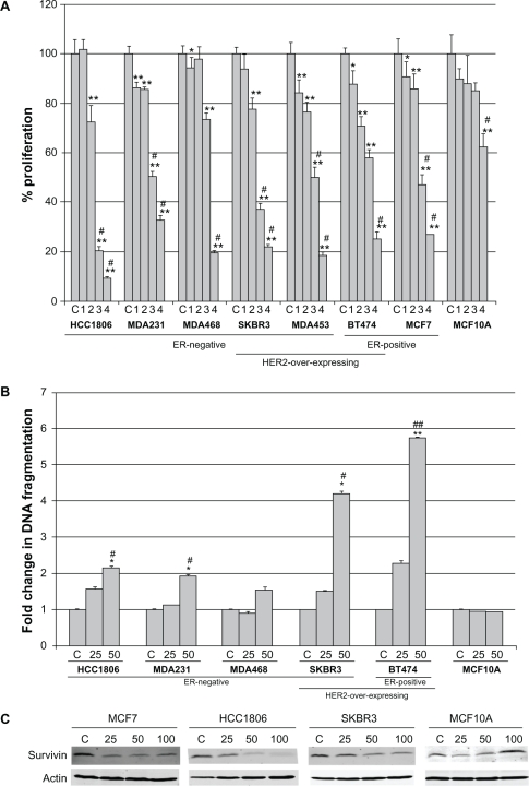
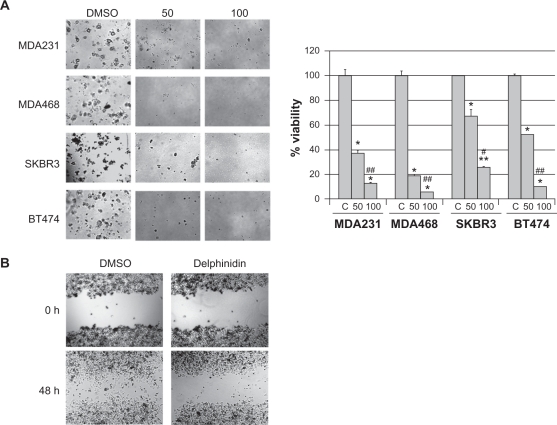
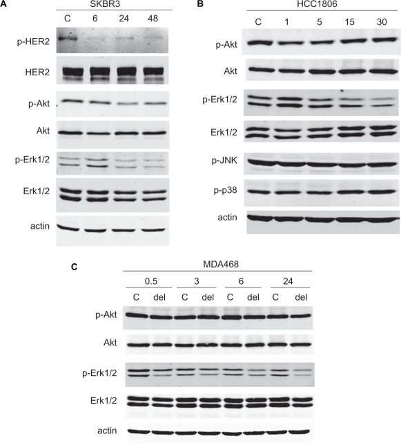
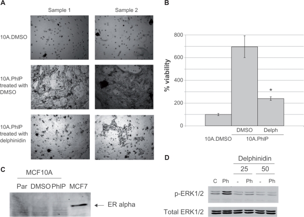
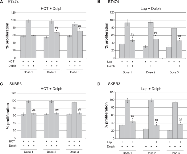
Delphinidin is a polyphenolic compound found in many brightly colored fruits and vegetables. Delphinidin is also the major bioactive component found in many dietary supplements that are currently consumed as complementary cancer medicine including pomegranate extract. The purpose of the current study was to determine the in vitro biological effects of delphinidin on established breast cancer cell lines of varying molecular subtypes in comparison to non-transformed breast epithelial cells. We examined cell proliferation, apoptosis, and growth inhibition in response to delphinidin using a tetrazolium salt-based assay, DNA fragmentation assay, and anchorage-independent growth assay. In comparison to vehicle control, delphinidin inhibited proliferation (P < 0.05), blocked anchorage-independent growth (P < 0.05), and induced apoptosis (P < 0.05) of ER-positive, triple negative, and HER2-overexpressing breast cancer cell lines with limited toxicity to non-transformed breast epithelial cells. MAPK signaling was partially reduced in triple negative cells and ER-negative chemically transformed MCF10A cells after treatment with delphinidin. In addition, delphinidin induced a significant level of apoptosis in HER2-overexpressing cells in association with reduced HER2 and MAPK signaling. Since delphinidin is often consumed as a complementary cancer medicine, the effect of delphinidin on response to specific HER2-targeted breast cancer therapies was examined by proliferation assay. Results of these drug combination studies suggested potential antagonism between delphinidin and HER2-directed treatments. In summary, the data presented here suggest that single agent delphinidin exhibits growth inhibitory activity in breast cancer cells of various molecular subtypes, but raise concerns regarding potential drug antagonism when used in combination with existing targeted therapies in HER2-overexpressing breast cancer.

Keywords: HER2; breast cancer; delphinidin; erbB2; triple negative.


Figures

Figure 1
Figure 1
Delphinidin inhibits proliferation of breast cancer cells with limited toxicity to non-transformed cells. (A) Breast cancer cells and non-transformed mammary epithelial cell line MCF10A were treated with 2-fold serial dilutions of delphinidin ranging from 12.5 μg/mL to 100 μg/mL. Control (C) cells were treated with DMSO at the volume found in the highest dose of delphinidin. After 6 days, proliferation was measured by MTS assay (Promega; Madison, WI). Treatments were done in 6 replicates, and experiments were performed on at least two separate occasions. Proliferation is shown as a percentage of DMSO control per cell line; error bars represent standard deviation between replicates. Statistical significance was determined across doses per cell line by single factor ANOVA; #P < 0.005. Statistical significance of each treatment versus control was determined per cell line by student’s t-test, *P < 0.05, **P < 0.005. (B) Breast cancer cells and MCF10A were treated with DMSO control or delphinidin at 25 μg/mL or 50 μg/mL. After 48 h, DNA fragmentation was measured using the Cell Death Detection ELISA kit (Roche; Indianapolis, IN). Each treatment group was done in duplicate. Fold change in DNA fragmentation is shown as a percentage of the control per cell line; error bars represent standard deviation between duplicates. Statistical significance of differences in DNA fragmentation was determined across doses per cell line by single factor ANOVA; #P < 0.05, ##P < 0.005. Statistical significance of each treatment versus control was determined per cell line by student’s t-test, *P < 0.05, **P < 0.005. (C) Cells were treated with 25, 50, or 100 μg/mL delphinidin for 48 h, or with DMSO at a volume equivalent to that in 100 μg/mL delphinidin as a control. Lysates were blotted for survivin anti-apoptotic protein; actin served as a loading control.
Figure 2
Figure 2
Delphinidin blocks anchorage-independent growth and migration of breast cancer cells. (A) Cells were plated in matrigel and treated with 50 μg/mL or 100 μg/mL delphinidin; control cells were treated with DMSO at the same volume as that found in the highest dose of delphinidin. Cells were maintained for 3–4 weeks, at which point photos were taken with an Olympus IX50 microscope at 4× magnification. Matrigel was then dissolved with dispase (5 mg/mL), and viable cells were counted by trypan blue exclusion. Cell survival is shown as a percentage of DMSO control cultures; error bars represent standard deviation between replicates. Statistical significance between doses including control was determined per cell line by single factor ANOVA; #P < 0.05, ##P < 0.005. Statistical significance between cell lines was determined by single factor ANOVA. Statistical significance of each treatment versus control was determined per cell line by student’s t-test, *P ≤ 0.05, **P < 0.005. (B) Confluent cultures of SKBR3 cells were scratched down the center with a pipet tip. Cells were then treated with 50 μg/mL delphinidin or DMSO control. Photos were taken with an Olympus IX50 microscope at 4× magnification at 0 h and 48 h after treatment.
Figure 3
Figure 3
Delphinidin inhibits HER2 and MAPK signaling in breast cancer cells. (A) SKBR3 cells were treated with 50 μg/mL delphinidin for 6, 24, or 48 h. (B) HCC1806 cells were treated with 50 μg/mL delphinidin for 1, 5, 15, or 30 min. Control cells were treated with DMSO at the volume found in this concentration of delphinidin for the longest time point (48 h in (A) or 30 min in (B)). Total protein lysates were immunoblotted for (A) phosphorylated and total HER2, Akt, and Erk1/2, or (B) p-Akt, total Akt, p-Erk1/2, total Erk1/2, p-JNK MAPK, and p-p38 MAPK; actin served as a loading control. (C) MDA468 cells were treated with 50 μg/mL delphinidin for 0.5, 3, 6, or 24 h. Control cells were treated with DMSO at the volume equivalent to that in 50 μg/mL delphinidin for 24 h. Lysates were blotted for phosphorylated and total Akt and Erk1/2; actin served as a loading control.
Figure 4
Figure 4
Delphinidin blocks survival of chemically transformed ER-negative MCF10A cells. MCF10A cells were maintained in DMSO or the carcinogen PhIP for 2 months, at which point cells were plated in matrigel. MCF10A.DMSO cells in matrigel were maintained in DMSO; MCF10A.PhIP cells in matrigel were either maintained in DMSO or 50 μg/mL delphinidin. Cells were maintained for 3–4 weeks, at which point (A) photos of duplicate cultures were taken for each treatment condition using an Olympus IX50 microscope at 4× magnification, and (B) matrigel was dissolved with dispase (5 mg/mL), and viable cells were counted by trypan blue exclusion. Cell survival is shown as a percentage of MCF10A.DMSO cells; error bars represent standard deviation between replicates. Statistical significance between delphinidin-treated MCF10A.PhIP cells versus control DMSO-treated MCF10A.PhIP cells was determined by student’s t-test; *P < 0.05. (C) MCF10A parental, DMSO control, and PhIP-transformed cells were Western blotted for expression of estrogen receptor using MCF-7 cell lysate as a positive control. (D) MCF10A cells were treated for 60 min with DMSO or 100 nM PhIP in the absence or presence of 25 μg/mL or 50 μg/mL delphinidin. Total protein lysates were immunoblotted for phosphorylated and total Erk1/2.
Figure 5
Figure 5
Delphinidin reduces anti-proliferative activity of HER2-targeted therapies: potential for drug antagonism. (A) BT474 cells were treated with 5, 10, or 20 μg/mL Herceptin (HCT), 6, 12, or 24 μg/mL delphinidin (delph), or combination HCT + delph. (B) BT474 cells were treated with 100, 200, or 400 nM lapatinib (lap), 6, 12, or 24 μg/mL delphinidin (delph), or combination lap + delph. (C) SKBR3 cells were treated with 5, 10, or 20 μg/mL Herceptin (HCT), 6, 12, or 24 μg/mL delphinidin (delph), or combination HCT + delph. (D) SKBR3 cells were treated with 100, 200, or 400 nM lapatinib (lap), 6, 12, or 24 μg/mL delphinidin (delph), or combination lap + delph. Control cells were treated with DMSO at the volume found in the highest concentration of delphinidin and/or lapatinib. After 6 d, MTS colorimetric assays were performed to measure cell proliferation. Notes: Values are expressed as a percentage of control DMSO group per line. Error bars represent standard deviation between six replicates. Statistical significance of differences between treatment groups was determined by ANOVA; ##P<0.005. Statistical significance of each treatment versus control was determined per cell line by student’s t-test; *P ≤ 0.05.

References

    1. Jemal A, Siegel R, Xu J, Ward E. Cancer statistics, 2010. CA Cancer J Clin. 2010;60:277–300. - PubMed
    1. Voduc KD, Cheang MC, Tyldesley S, Gelmon K, et al. Breast cancer subtypes and the risk of local and regional relapse. J Clin Oncol. 2010;28:1684–91. - PubMed
    1. Millar EK, Graham PH, O’Toole SA, McNeil CM, et al. Prediction of local recurrence, distant metastases, and death after breast-conserving therapy in early-stage invasive breast cancer using a five-biomarker panel. J Clin Oncol. 2009;27:4701–8. - PubMed
    1. Nguyen PL, Taghian AG, Katz MS, Niemierko A, et al. Breast cancer subtype approximated by estrogen receptor, progesterone receptor, and HER-2 is associated with local and distant recurrence after breast-conserving therapy. J Clin Oncol. 2008;26:2373–8. - PubMed
    1. Haffty BG, Yang Q, Reiss M, Kearney T, et al. Locoregional relapse and distant metastasis in conservatively managed triple negative early-stage breast cancer. J Clin Oncol. 2006;24:5652–7. - PubMed