. 2011:2011:591356.
doi: 10.1093/ecam/neq053. Epub 2011 Apr 14.

Quercetin and cancer chemoprevention

Affiliations

Quercetin and cancer chemoprevention

Lara Gibellini et al. Evid Based Complement Alternat Med. 2011.

Abstract

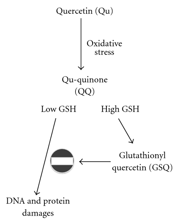
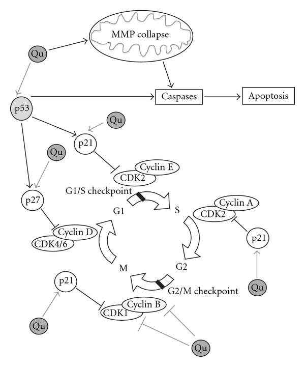
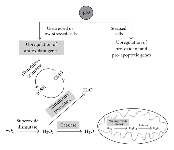
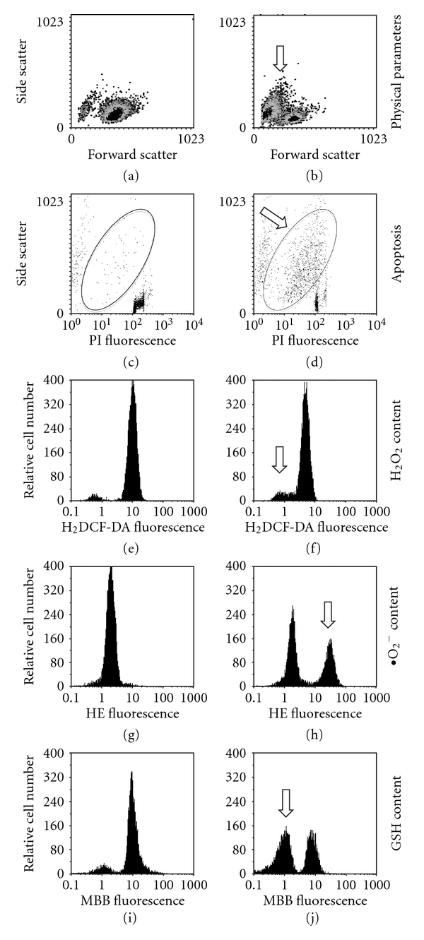
Several molecules present in the diet, including flavonoids, can inhibit the growth of cancer cells with an ability to act as "chemopreventers". Their cancer-preventive effects have been attributed to various mechanisms, including the induction of cell-cycle arrest and/or apoptosis as well as the antioxidant functions. The antioxidant activity of chemopreventers has recently received a great interest, essentially because oxidative stress participates in the initiation and progression of different pathological conditions, including cancer. Since antioxidants are capable of preventing oxidative damage, the wide use of natural food-derived antioxidants is receiving greater attention as potential anti-carcinogens. Among flavonoids, quercetin (Qu) is considered an excellent free-radical scavenging antioxidant, even if such an activity strongly depends on the intracellular availability of reduced glutathione. Apart from antioxidant activity, Qu also exerts a direct, pro-apoptotic effect in tumor cells, and can indeed block the growth of several human cancer cell lines at different phases of the cell cycle. Both these effects have been documented in a wide variety of cellular models as well as in animal models. The high toxicity exerted by Qu on cancer cells perfectly matches with the almost total absence of any damages for normal, non-transformed cells. In this review we discuss the molecular mechanisms that are based on the biological effects of Qu, and their relevance for human health.


Figures

Figure 1
Figure 1
Antioxidant and pro-oxidant effects of Qu in the presence of low and high levels of reduced GSH. The antioxidant and pro-oxidant effects of Qu strongly depend upon the availability of intracellular reduced GSH. During an oxidative stress, in the presence of peroxidases, Qu reacts with H2O2 to form a semiquinone radical that is rapidly oxidized to QQ. QQ has a pro-oxidant effect; its high reactivity towards protein thiols and DNA leads to cell damage and cytotoxicity. QQ is also highly reactive towards thiols, and preferentially reacts with GSH to form relatively stable protein-oxidized Qu adducts such as 6-GSQ and 8-GSQ. The reversibility of this reaction allows the continuous dissociation of GSQ into GSH and QQ. In the presence of high GSH concentrations, QQ reacts with GSH to form GSQ again, and QQ cannot exert its cytotoxic effects, whereas when low levels of GSH are present, QQ reacts with protein thiols, thus leading to cellular damage.
Figure 2
Figure 2
Effects of Qu on cell cycle. Qu is able to regulate cell cycle by directly binding several molecular targets and, depending on the cell type and tumor origin, it blocks the cell cycle at G2/M or at the G1/S transition. At the G1/S transition, Qu blocks cell-cycle progression through the up-regulation of p21 and p27 and p53. p21 exerts an inhibitory activity on several CDKs. In particular, p21 inhibits CDK2-cyclin E, with the consequent inhibition of CDK2-dependent phosphorylation of pRb and the sequestration of E2F1, thus inhibiting gene transcription induced by E2F1 and progression into and through S phase. p21 also inhibits CDK2-cyclin A and CDK1-cyclin B, which are essential for progression through S phase and G2, respectively. p27 exerts several effects on cell cycle, but only under certain conditions it can inhibit the complexes CDK4-cyclin D and CDK6-cyclin D. The tumor suppressor p53, once activated, can induce several different cellular responses, including growth arrest and apoptosis. Growth arrest is essentially elicited through the up-regulation of the genes that encode for inhibitors of cell-cycle progression, including p21 and p27. In different cellular models, Qu stabilizes p53 both at mRNA and protein levels. Apart from blocking cell growth through the direct action on key modulators of cell cycle, Qu is able to induce apoptosis via mitochondrial pathway: indeed, Qu can disrupt MMP, which in turn provokes the release of cytochrome c in the cytoplasm, a phenomenon that activates multiple caspases, such as caspase-3 and -7.
Figure 3
Figure 3
Effects of p53 in the intracellular oxidant system. According to a new model of p53-dependent regulation of ROS, p53 acts on the intracellular antioxidant system in both unstressed and low-stressed cells through the up-regulation of a series of genes (indicated in light gray boxes), including Gpx1, Mn-SOD2 and catalase. GPX catalyzes the oxidation of GSH into GSSG; Mn-SOD2 is localized in mitochondria and catalyzes the dismutation of superoxide (O2 ) into oxygen and hydrogen peroxide (H2O2), whereas catalase catalyzes the decomposition of H2O2 to water and oxygen. When high levels of stress are present, p53 induces the transcription of several pro-oxidant as well as pro-apoptotic genes, such as bax (in dark gray circle), with the consequent release of cytochrome c from mitochondria, activation of caspases and induction of apoptosis.
Figure 4
Figure 4
Changes in cell viability, apoptosis and content of H2O2, O2 and GSH in U937 tumor cell line treated for 24 h with Qu 100 μM. U937 cells treated with 100 μM Qu were separately stained with four fluorescent dyes: propidium iodide (PI), 2′,7′-dichlorodihydrofluorescein diacetate (H2-DCFDA), hydroethidine (HE) and monobromobimane (MBB) for the quantification of nuclear DNA, intracellular H2O2, intracellular O2 and intracellular GSH content, respectively. Cells were then analyzed by flow cytometry. In (a) and (b) the physical parameters (identified by forward and side scatter for cell dimension and granularity, resp.) of the cells under investigation are represented. Treating cells with 100 μM Qu causes an increase in the number of cells with reduced forward scatter and increased side scatter, typical of apoptosis (indicated in b by an arrow). (c) and (d) Changes in DNA content (PI fluorescence) and side scatter in control and Qu-treated cells. In the ellipse, cells with hypodyploid DNA content and increased side scatter (i.e., those apoptotic) are present, and increase after treatment with Qu. (e) and (f) Show intracellular H2O2 content in both control and treated cells. Qu causes a shift to the left of the fluorescence peak (see, in (e), the histogram shift in relation to the fix position of the red bars) indicating a small reduction in H2O2 content, likely because of the concomitant increase in O2 . Qu also causes a small change in the percentage of cells that do not bind the dye, that is, those undergoing apoptosis (arrow), which are those evidenced in (d). (g) and (h) Represent intracellular O2 content in the absence or in the presence of 100 μM Qu. Treating cells with Qu significantly increases the amount of cells with high HE fluorescence (arrow), which represents an increase of intracellular O2 content. (i) and (j) Show GSH content in the absence or after treatment with Qu 100 μM. In the presence of Qu, MBB fluorescence decreases in a consistent number of cells (arrow), indicating that Qu is able to deplete intracellular GSH.

References

    1. Kennedy DA, Hart J, Seely D. Cost effectiveness of natural health products: a systematic review of randomized clinical trials. Evidence-Based Complementary and Alternative Medicine. 2009;6(3):297–304. - PMC - PubMed
    1. Danaei G, Vander Hoorn S, Lopez AD, Murray CJL, Ezzati M. Causes of cancer in the world: comparative risk assessment of nine behavioural and environmental risk factors. Lancet. 2005;366(9499):1784–1793. - PubMed
    1. Knekt P, Kumpulainen J, Järvinen R, et al. Flavonoid intake and risk of chronic diseases. American Journal of Clinical Nutrition. 2002;76(3):560–568. - PubMed
    1. Manson MM. Cancer prevention—the potential for diet to modulate molecular signalling. Trends in Molecular Medicine. 2003;9(1):11–18. - PubMed
    1. Van Duijnhoven FJB, Bueno-De-Mesquita HB, Ferrari P, et al. Fruit, vegetables, and colorectal cancer risk: the European Prospective Investigation into Cancer and Nutrition. American Journal of Clinical Nutrition. 2009;89(5):1441–1452. - PubMed