Review
. 2011 Sep 14;111(9):5768-83.
doi: 10.1021/cr100006x. Epub 2011 Jul 27.

Reactivity of thioredoxin as a protein thiol-disulfide oxidoreductase

Affiliations
Review

Reactivity of thioredoxin as a protein thiol-disulfide oxidoreductase

Zhiyong Cheng et al. Chem Rev. .
No abstract available


Figures

Figure 1
Figure 1
A schematic cartoon view of a Trx-catalyzed disulfide reduction. A number of factors are involved in the regulation of Trx activity, including the amino acid residues spanning the redox active CXXC motif, molecular interaction (e.g., electrostatic force), acid/base catalysts (B1 and B2), conformational adjustment to orientate the reactive groups to facilitate reaction, and hydrophobic microenvironments formed after substrate binding (indicated in pink). Taking EcTrx as an example, the reaction of Trx with target disulfide proteins includes binding (panel A to B), the first nucleophilic attack of Cys32-S- (somewhat exposed to the solvent, panel A) on the target disulfide to form an intermolecular mixed disulfide intermediate (panel B to panel C), followed by a second nucleophilic attack of Cys35-S (buried in EcTrx) on the mixed disulfide (panel C), with the generation of Trx-S2 and the reduced protein (panel D). Binding of the protein substrate to Trx could create a more apolar environment for Cys32 and place it in proximity to acid/base catalysts. However, the identity of acid/base catalysts still remain to be established, although they are critical for the ionization of thiol groups in CXXC motif for nucleophilic attack.
Figure 2
Figure 2
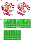
The structure of EcTrx and conserved amino acid residues in the Trx family. (A) and (B) are the ribbon diagrams of oxidized and reduced EcTrx, respectively. They show the spatial distances between Trp28 and Cys32, and between Trp28 and Asp26. The color of the backbone labels the secondary structures, where red indicates alpha-helices, yellow indicates beta-sheets, and gray indicates turns or coils. The atom coordinates of proteins are obtained from the PDB, and the figures of protein structures were generated using RasMol. (C-E) show the conserved residues with sequence alignments of 515 Trx sequences (C), 495 DsbA sequences (D), and 382 PDI-α protein sequences (E) from the Conserved Domain Database of NCBI (note that only the more conserved residues are shown). For each panel,(C) - (E), the top portion shows the relative conservation of residues at each conserved position, the middle shows the consensus sequence, and the bottom portion shows the Master Sequence of each class (2TRX_A, 1DSB_A, and 1MEK, respectively). The Master Sequence is that of a real protein, and is the sequence to which all other sequences in the Conserved Domain (CD) alignment are related pair-wise. The Consensus Sequence for a CD contains, at each position, the most frequently occurring amino acid at that position in the alignment of the CD. The relative heights of the green boxes show the residue (in black) having the highest fraction among all the aligned sequences. The color gradation from green to red indicates a decreased conservation of the corresponding residue. From the alignment of the Master Sequences, we can see that the two Cys residues of the three representative proteins (32 and 35 of 2TrxA, 30 and 33 of 1DsbA, and 36 and 39 of 1MEK) are highly conserved in their corresponding families. The alignment also suggests a proximal proline is almost completely conserved in these families (Pro76 in EcTrx, PDB: 2TrxA; Pro151 in EcDsbA, PDB: 1DsbA; and Pro83 in human PDI, PDB: 1MEK). (F) Spatial distance of active site residues Cys32 and Cys35 from Pro76 in EcTrx. (G) Spatial distance between Asp26 and Cys35, and between Pro76 and Cys35 of EcTrx.
Figure 2
Figure 2
The structure of EcTrx and conserved amino acid residues in the Trx family. (A) and (B) are the ribbon diagrams of oxidized and reduced EcTrx, respectively. They show the spatial distances between Trp28 and Cys32, and between Trp28 and Asp26. The color of the backbone labels the secondary structures, where red indicates alpha-helices, yellow indicates beta-sheets, and gray indicates turns or coils. The atom coordinates of proteins are obtained from the PDB, and the figures of protein structures were generated using RasMol. (C-E) show the conserved residues with sequence alignments of 515 Trx sequences (C), 495 DsbA sequences (D), and 382 PDI-α protein sequences (E) from the Conserved Domain Database of NCBI (note that only the more conserved residues are shown). For each panel,(C) - (E), the top portion shows the relative conservation of residues at each conserved position, the middle shows the consensus sequence, and the bottom portion shows the Master Sequence of each class (2TRX_A, 1DSB_A, and 1MEK, respectively). The Master Sequence is that of a real protein, and is the sequence to which all other sequences in the Conserved Domain (CD) alignment are related pair-wise. The Consensus Sequence for a CD contains, at each position, the most frequently occurring amino acid at that position in the alignment of the CD. The relative heights of the green boxes show the residue (in black) having the highest fraction among all the aligned sequences. The color gradation from green to red indicates a decreased conservation of the corresponding residue. From the alignment of the Master Sequences, we can see that the two Cys residues of the three representative proteins (32 and 35 of 2TrxA, 30 and 33 of 1DsbA, and 36 and 39 of 1MEK) are highly conserved in their corresponding families. The alignment also suggests a proximal proline is almost completely conserved in these families (Pro76 in EcTrx, PDB: 2TrxA; Pro151 in EcDsbA, PDB: 1DsbA; and Pro83 in human PDI, PDB: 1MEK). (F) Spatial distance of active site residues Cys32 and Cys35 from Pro76 in EcTrx. (G) Spatial distance between Asp26 and Cys35, and between Pro76 and Cys35 of EcTrx.
Figure 3
Figure 3
Plot of the change in A340 as the reaction comes to equilibrium vs. the initial ratios of reactants. The cross-over point where the change in A340 is zero gives the Keq value. Shown are data for determining Keq for DmTrx2 (pH 7.0, 0.1 M potassium phosphate buffer containing 0.3 mM EDTA). The initial ratio of [DmTrx(SH)2]/[DmTrx(S)2] was kept constant at 5. The initial concentrations of NADPH and NADP+ were varied systematically to give the ratios of [NADP]/[NADPH] from 8 to 16, but the sum of the concentrations of NADP + NADPH was maintained at 220 μM. The initial ratios of [NADP] [DmTrx(SH)2]/[NADPH] [DmTrx(S)2] ranged from 40 to 80 accordingly. Further details concerning the calculation and related examples can be found in reference 66. Reprinted with permission from Reference 66. Copyright 2007 American Chemical Society.
Figure 4
Figure 4
A schematic view of a proposed catalytic tetrad composed of Asp65, Trp35, the redox-active disulfide (Cys36-Cys39), and the protonated buried Asp30 in CrTrxh. The catalytic tetrad provides both base catalysis toward Cys36-SH, provided by an aromatic side chain of Trp35, and acid catalysis toward Cys39-SH, provided by Asp30. Adapted with permission from Reference 44. Copyright 1998 Wiley-Blackwell.
Figure 5
Figure 5
A schematic hypothesis of Pro151 in DsbA in aiding the shuttling of a proton in the cleavage of DsbA-substrate mixed disulfide.
Figure 6
Figure 6
Electrostatic properties of thioredoxins. Charge distribution on molecular surfaces of E. coli wild type Trx (a), spinach chloroplast Trx-f (b), E30K/L94K E. coli Trx (c), and spinach chloroplast Trx-m (d), as calculated with GRASP. Electrostatic potentials at the protein surface and superimposed isopotential shells (calculated at ±2 kT) are colored as follows: red as negative and blue as positive. The active site regions of all proteins are located at the center of each image. An S illustrates the position of the accessible sulfur atom of Cys-32. Reprinted with permission from Reference 122. Copyright 1998 American Society for Biochemistry and Molecular Biology.
Figure 7
Figure 7
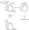
A scheme showing that an intramolecular disulfide formed between the catalytically inactive thiols (i.e., Cys62–Cys69 disulfide of human Trx) impaired Trx activity by disrupting Trx–target protein interactions. Due to this impairment, the half-life of the oxidized Trx becomes prolonged, for which Trx activity can be transiently inhibited under conditions of redox signaling or oxidative stress, and allows more time for the sensing and transmission of oxidative signals. Adapted with permission from Reference 135. Copyright 2004 Oxford University Press.

References

    1. Jensen KS, Hansen RE, Winther JR. Antioxid Redox Signal. 2009;11:1047. - PubMed
    1. Hatahet F, Ruddock LW. Antioxid Redox Signal. 2009;11:2807. - PubMed
    1. Jeong W, Yoon HW, Lee SR, Rhee SG. J Biol Chem. 2004;279:3142. - PubMed
    1. Powis G, Montfort WR. Annu Rev Pharmacol Toxicol. 2001;41:261. - PubMed
    1. Lillig CH, Holmgren A. Antioxid Redox Signal. 2007;9:25. - PubMed

Publication types

MeSH terms

Substances