. 2011 Oct;85(19):10310-8.
doi: 10.1128/JVI.05252-11. Epub 2011 Jul 27.

Creation of a recombinant Rift Valley fever virus with a two-segmented genome

Affiliations

Creation of a recombinant Rift Valley fever virus with a two-segmented genome

Benjamin Brennan et al. J Virol. 2011 Oct.

Abstract

Rift Valley fever virus (RVFV; family Bunyaviridae) is a clinically important, mosquito-borne pathogen of both livestock and humans, which is found mainly in sub-Saharan Africa and the Arabian Peninsula. RVFV has a trisegmented single-stranded RNA (ssRNA) genome. The L and M segments are negative sense and encode the L protein (viral polymerase) on the L segment and the virion glycoproteins Gn and Gc as well as two other proteins, NSm and 78K, on the M segment. The S segment uses an ambisense coding strategy to express the nucleocapsid protein, N, and the nonstructural protein, NSs. Both the NSs and NSm proteins are dispensable for virus growth in tissue culture. Using reverse genetics, we generated a recombinant virus, designated r2segMP12, containing a two-segmented genome in which the NSs coding sequence was replaced with that for the Gn and Gc precursor. Thus, r2segMP12 lacks an M segment, and although it was attenuated in comparison to the three-segmented parental virus in both mammalian and insect cell cultures, it was genetically stable over multiple passages. We further show that the virus can stably maintain an M-like RNA segment encoding the enhanced green fluorescent protein gene. The implications of these findings for RVFV genome packaging and the potential to develop multivalent live-attenuated vaccines are discussed.


Figures

Fig. 1.
Fig. 1.
Comparison of plaque sizes of rMP12, r2segMP12, and rMP12-delNSs:eGFP-delNSm on BHK-21 cells. Cell monolayers were fixed 96 h p.i. with 4% paraformaldehyde and stained with Giemsa solution.
Fig. 2.
Fig. 2.
Analysis of S segment RNA from BHK-21 cells infected with parental (rMP12) or recombinant viruses (r2segMP12). (A) Schematic depiction of the parental S segment, the engineered S segment, and the sites at which oligonucleotides (α, β, and ψ) anneal to the genomic segments. (B and C) RT-PCR was used to confirm that the two-segmented virus had an S segment that encoded the viral glycoproteins. BHK-21 cells were infected with rMP12 or r2segMP12 viruses at an MOI of 1. Total cellular RNA was extracted from the infected cells 48 h p.i., and S-segment RT-PCR was performed. Amplified DNA products from PCRs on the viral cDNAs using oligonucleotides β & ψ (B) and α & β (C) are shown.
Fig. 3.
Fig. 3.
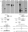
(Top) Analysis of viral RNAs synthesized in infected cells. BHK-21 cells were infected with rMP12 or r2segMP12 (MOI = 1), and at 48 h p.i. total cell RNA was isolated. Northern blotting was performed using DIG-labeled probes complementary to N (A), Gn (B), L (D) or NSs (F) coding sequences in the viral genomic (−) sense RNA, or Gn (C) or N (E) coding sequences in the viral anti-genomic (+) sense RNA. The sizes of the RNA species are indicated on the left. (Bottom) A schematic of the viral genome structures of rMP12 and r2segMP12 and the binding regions for the DIG-labeled probes described for panel A. (−/+) indicates whether the probe detects viral genomic (−) or anti-genomic (+) sense RNA based on the genome structure of rMP12. Double-ended arrows represent genomic replication, and single-ended arrows represent transcription of the genome. (▪) indicates 5′ cap structure on messenger RNAs.
Fig. 4.
Fig. 4.
Growth properties of recombinant viruses. Viral growth curves were monitored in BHK-21 (A), Vero E6 (B), and C6/36 (C) cells following infection with rMP12, r2segMP12, or rMP12-delNSs:eGFP-delNSm (MOI = 1). Viruses were harvested at the time points indicated and titrated by plaque assay. Graphs are presented for one representative experiment. (D) Western blot analysis of S segment-encoded proteins from rMP12, r2segMP12, and rMP12-delNSs:eGFP-delNSm-infected cells. Cell extracts were prepared from the growth curve samples at the time points indicated, proteins fractionated on 4 to 12% NuPage gels, and blots were probed with anti-N, anti-NSs, and anti-tubulin antibodies as indicated.
Fig. 5.
Fig. 5.
Western blot analysis of Gn glycoprotein and N protein produced during rMP12, r2segMP12, and rMP12-delNSs:eGFP-delNSm infection. Infected cell extracts were harvested at the time points indicated, and protein blots probed with anti-N and anti-Gn antibodies. Intensities of the bands for N and Gn were measured using a luminescent image analyzer (Fujifilm) and a ratio estimated at the 24-h-p.i. time point (▪).
Fig. 6.
Fig. 6.
Effect of multiplicity of infection on viral titer. BHK-21 cells were infected with rMP12 or r2segMP12 at a range of multiplicities from 0.0005 to 5 PFU/cell. Viral supernatants were harvested 72 h p.i. and titrated by plaque assay. The results are presented for one representative experiment.
Fig. 7.
Fig. 7.
Serial passage of rMP12 and r2segMP12 in BHK-21 cells. (A) Cell extracts were prepared at different passages as indicated, and protein blots were probed with anti-N, anti-NSs, and anti-tubulin antibodies. (B) Virus supernatant was also harvested at the passages indicated and titrated by plaque assay on BHK-21 cells.
Fig. 8.
Fig. 8.
Characterization of recombinant virus containing an M-like RNA segment expressing eGFP. (A) eGFP autofluorescence in transfected cells (Rescue) or cells infected with r2segMP12:MeGFPM after 1 (P1), 2 (P2), or 3 (P3) passages. (B) Comparison of r2segMP12 and r2segMP12:MeGFPM plaque sizes on BHK-21 cells. Cell monolayers were fixed 96 h p.i. with 4% paraformaldehyde and stained with Giemsa solution. (C) RNA analysis following serial passage of r2segMP12:MeGFPM in BHK-21 cells. BHK-21 cells were infected with rMP12 or r2segMP12:MeGFPM viruses either directly from the rescue cells (R) or at different passage levels (P1, P2, or P3 as indicated), and at 48 h p.i., total cell RNA was isolated. Northern blotting was performed using DIG-labeled probes complementary to NSm or eGFP coding sequences in the viral genomic RNA (−) and N or Gn coding sequences in the viral anti-genomic RNA (+). The sizes of the RNA species are indicated on the left.

References

    1. Billecocq A., et al. 2008. RNA polymerase I-mediated expression of viral RNA for the rescue of infectious virulent and avirulent Rift Valley fever viruses. Virology 378:377–384 - PMC - PubMed
    1. Billecocq A., et al. 2004. NSs protein of Rift Valley fever virus blocks interferon production by inhibiting host gene transcription. J. Virol. 78:9798–9806 - PMC - PubMed
    1. Bird B. H., et al. 2008. Rift valley fever virus lacking the NSs and NSm genes is highly attenuated, confers protective immunity from virulent virus challenge, and allows for differential identification of infected and vaccinated animals. J. Virol. 82:2681–2691 - PMC - PubMed
    1. Bird B. H., Albariño C. G., Nichol S. T. 2007. Rift Valley fever virus lacking NSm proteins retains high virulence in vivo and may provide a model of human delayed onset neurologic disease. Virology 362:10–15 - PubMed
    1. Bouloy M., Flick R. 2009. Reverse genetics technology for Rift Valley fever virus: current and future applications for the development of therapeutics and vaccines. Antiviral Res. 84:101–118 - PMC - PubMed

Publication types

LinkOut - more resources