. 2011 Oct;85(19):10144-53.
doi: 10.1128/JVI.05035-11. Epub 2011 Jul 27.

Epstein-Barr virus latent membrane protein 1 (LMP1) C-terminal-activating region 3 contributes to LMP1-mediated cellular migration via its interaction with Ubc9

Affiliations

Epstein-Barr virus latent membrane protein 1 (LMP1) C-terminal-activating region 3 contributes to LMP1-mediated cellular migration via its interaction with Ubc9

Gretchen L Bentz et al. J Virol. 2011 Oct.

Abstract

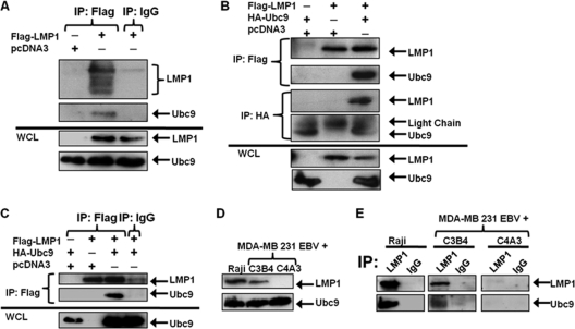
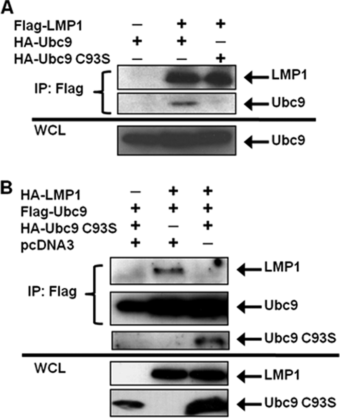
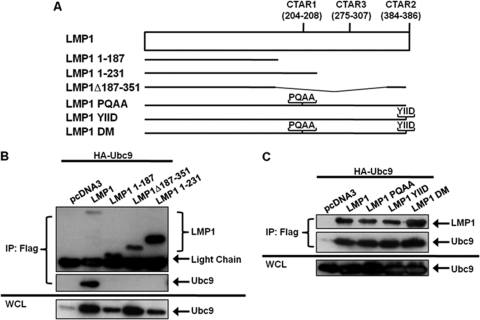
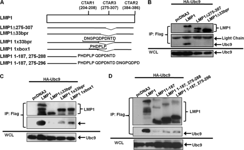
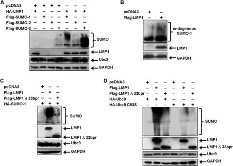
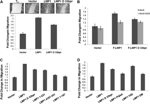
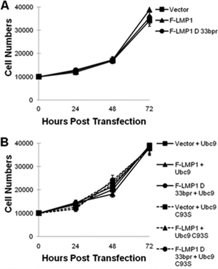
Epstein-Barr virus (EBV) latent membrane protein 1 (LMP1), the principal viral oncoprotein and a member of the tumor necrosis factor receptor superfamily, is a constitutively active membrane signaling protein that regulates multiple signal transduction pathways via its C-terminal-activating region 1 (CTAR1) and CTAR2, and also the less-studied CTAR3. Because protein sumoylation among other posttranslational modifications may regulate many signaling pathways induced by LMP1, we investigated whether during EBV latency LMP1 regulates sumoylation processes that control cellular activation and cellular responses. By immunoprecipitation experiments, we show that LMP1 interacts with Ubc9, the single reported SUMO-conjugating enzyme. Requirements for LMP1-Ubc9 interactions include enzymatically active Ubc9: expression of inactive Ubc9 (Ubc9 C93S) inhibited the LMP1-Ubc9 interaction. LMP1 CTAR3, but not CTAR1 and CTAR2, participated in the LMP1-Ubc9 interaction, and amino acid sequences found in CTAR3, including the JAK-interacting motif, contributed to this interaction. Furthermore, LMP1 expression coincided with increased sumoylation of cellular proteins, and disruption of the Ubc9-LMP1 CTAR3 interaction almost completely abrogated LMP1-induced protein sumoylation, suggesting that this interaction promotes the sumoylation of downstream targets. Additional consequences of the disruption of the LMP1 CTAR3-Ubc9 interaction revealed effects on cellular migration, a hallmark of oncogenesis. Together, these data demonstrate that LMP1 CTAR3 does in fact function in intracellular signaling and leads to biological effects. We propose that LMP1, by interaction with Ubc9, modulates sumoylation processes, which regulate signal transduction pathways that affect phenotypic changes associated with oncogenesis.


Figures

Fig. 1.
Fig. 1.
EBV LMP1 interacts with Ubc9. (A) 293T cells were transfected with FLAG-LMP1 or vector control and cultured for 48 h. Cell lysates were collected and immunoprecipitations (IP) were performed with FLAG or isotype control antibodies. Western blot analyses of the immunoprecipitates, and whole-cell lysates (WCL; 10 μg, or 2% of total lysates) were obtained for detection of FLAG-LMP1 and endogenous Ubc9. (B) 293T cells were transfected with FLAG-LMP1 and/or HA-Ubc9 and cultured for 48 h. Cell lysates were collected, and immunoprecipitations were performed with FLAG and HA antibodies. Western blot analyses were performed to detect FLAG-LMP1 and HA-Ubc9 expression. (C) 293T cells were transfected with FLAG-LMP1 and/or HA-Ubc9 and cultured for 48 h. Cell lysates were collected, and immunoprecipitations were performed with FLAG antibodies or isotype control antibodies. Western blot analyses were performed to detect FLAG-LMP1 and HA-Ubc9 expression. (D) WCL of Raji cells and MDA-MB 231 EBV+ cell clones C3B4 and C4A3 were subjected to Western blot analyses for detection of endogenous LMP1 and Ubc9. (E) Cell lysates were collected from Raji cells and MDA-MB 231 EBV+ cell clones C3B4 and C4A3. One milligram of total protein from each cell lysate was subjected to immunoprecipitations with LMP1 and IgG isotype control antibodies. Western blot analyses were performed to detect LMP1 and Ubc9 expression.
Fig. 2.
Fig. 2.
EBV LMP1 interacts only with enzymatically active Ubc9. (A) 293T cells were transfected with FLAG-LMP1 and HA-Ubc9 or HA-Ubc9 C93S. (B) 293T cells were transfected with FLAG-Ubc9, HA-LMP1, and increasing amounts of HA-Ubc9 C93S. At 48 h after transfection, cell lysates were harvested and immunoprecipitations (IP) were performed with FLAG antibodies. Western blot analyses of the immunoprecipitates and WCL were used to detect FLAG and HA expression.
Fig. 3.
Fig. 3.
LMP1 CTAR1 and CTAR2 are not critical for the LMP1-Ubc9 interaction. (A) Diagrammatic alignment of wild-type LMP1 and selected LMP1 mutants. LMP 1–187 is missing both CTAR1 and CTAR2. LMP1 1–231 only contains CTAR1. LMP1 Δ187–351 only contains CTAR2. LMP1 PQAA, YIID, and DM are point mutants with inactive CTAR1 and/or CTAR2. (B and C) 293T cells were transfected with FLAG-LMP1 or the indicated FLAG-LMP1 mutants and HA-Ubc9. Cell lysates were harvested 48 h after transfection, and immunoprecipitations were performed with FLAG antibodies. Western blot analyses of the immunoprecipitates and WCL were used to detect FLAG-LMP1 and HA-Ubc9 expression.
Fig. 4.
Fig. 4.
LMP1 CTAR3 is required for its interaction with Ubc9. (A) Diagrammatic alignment of LMP1 mutants. LMP1 Δ275–307 was constructed to delete both JAK3-binding motifs. LMP1Δ33bpr is missing the 33-bp repeats, which includes the JAK3-binding motifs. LMP1 1xbox 1 and LMP1 1x33bor are reconstituted LMP1Δ33bpr mutants. (B to D) 293T cells were transfected with HA-Ubc9 and FLAG-LMP1 or the indicated FLAG-LMP1 mutant. Cell lysates were harvested 48 h after transfection, and immunoprecipitations were performed with FLAG antibodies. Western blot analyses of the immunoprecipitates and WCL were performed to determine FLAG and HA expression.
Fig. 5.
Fig. 5.
LMP1 induces the sumoylation of cellular proteins. (A) 293T cells were transfected with vector control or HA-LMP1 and FLAG-SUMO-1, FLAG-SUMO-2, or FLAG-SUMO-2. (B) 293T cells were transfected with vector control or FLAG-LMP1. (C) 293T cells were transfected with vector control, FLAG-LMP1, or FLAG-LMP1Δ33bpr and HA-SUMO-1. (D) 293T cells were transfected with HA-Ubc9 or HA-Ubc9 C93S along with HA-SUMO-1 and vector control, FLAG-LMP1, or FLAG-LMP1Δ33bpr. At 48 h after transfection, whole-cell lysates were collected and Western blot analyses were performed for SUMO-1, FLAG, HA, Ubc9, and GAPDH expression.
Fig. 6.
Fig. 6.
Inhibition of the Ubc9-LMP1 CTAR3 interaction decreases cellular migration. (A) MDA-MB 231 EBV+ clone C4A3 cells were transfected to stably express vector control, HA-LMP1, or HA-LMP1Δ33bpr. Following selection, cells were plated and grown to confluence. Confluent cell layers were scratched with a pipette tip and washed, and fresh RPMI was added to wells. After 24 h, medium was removed and cells were stained and fixed in a crystal violet and ethanol solution. Multiple images were captured along the scratch, and the wound width was calculated (2, 4). Representative images are shown, and the fold change in migration was determined. (B to D) 293 cells were plated and transfected as indicated. Confluent cell layers were scratched with a pipette tip and washed, and fresh 1% DMEM was added to wells. After 18 h, medium was removed and cells were stained and fixed in crystal violet-ethanol. The wound width was calculated (2, 4), and the fold change in migration was determined. Data are the means ± standard deviations for experiments performed in triplicate.
Fig. 7.
Fig. 7.
The LMP1-Ubc9 interaction does not affect cellular growth. (A) A total of 1 × 10⁴ 293 cells were plated and transfected with vector control, FLAG-LMP1, or FLAG-LMP1Δ33bpr. (B) A total of 1 × 10⁴ 293 cells were plated and transfected with HA-Ubc9 or HA-Ubc9 C93S and either the vector control, FLAG-LMP1, or FLAG-LMP1Δ33bpr. Cells and supernatant fluids were collected at 24, 48, 72, and 96 h after transfection, and the total numbers of cells were determined.

References

    1. Adamson A. L., Kenney S. 2001. Epstein-Barr virus immediate-early protein BZLF1 is SUMO-1 modified and disrupts promyelocytic leukemia bodies. J. Virol. 75:2388–2399 - PMC - PubMed
    1. Albuquerque M. L., Waters C. M., Savla U., Schnaper H. W., Flozak A. S. 2000. Shear stress enhances human endothelial cell wound closure in vitro. Am. J. Physiol. Heart Circ. Physiol. 279:H293–H302 - PubMed
    1. Aviel S., Winberg G., Massucci M., Ciechanover A. 2000. Degradation of the Epstein-Barr virus latent membrane protein 1 (LMP1) by the ubiquitin-proteasome pathway. Targeting via ubiquitination of the N-terminal residue. J. Biol. Chem. 275:23491–23499 - PubMed
    1. Bentz G. L., Yurochko A. D. 2008. Human CMV infection of endothelial cells induces an angiogenic response through viral binding to EGF receptor and β1 and β3 integrins. Proc. Natl. Acad. Sci. U. S. A. 105:5531–5536 - PMC - PubMed
    1. Brennan P., Floettmann J. E., Mehl A., Jones M., Rowe M. 2001. Mechanism of action of a novel latent membrane protein-1 dominant negative. J. Biol. Chem. 276:1195–1203 - PubMed

Publication types

Substances