. 2011 Sep;22(18):3533-40.
doi: 10.1091/mbc.E11-06-0483. Epub 2011 Jul 27.

GSK3β inactivation induces apoptosis of leukemia cells by repressing the function of c-Myb

Affiliations

GSK3β inactivation induces apoptosis of leukemia cells by repressing the function of c-Myb

Fangfang Zhou et al. Mol Biol Cell. 2011 Sep.

Abstract

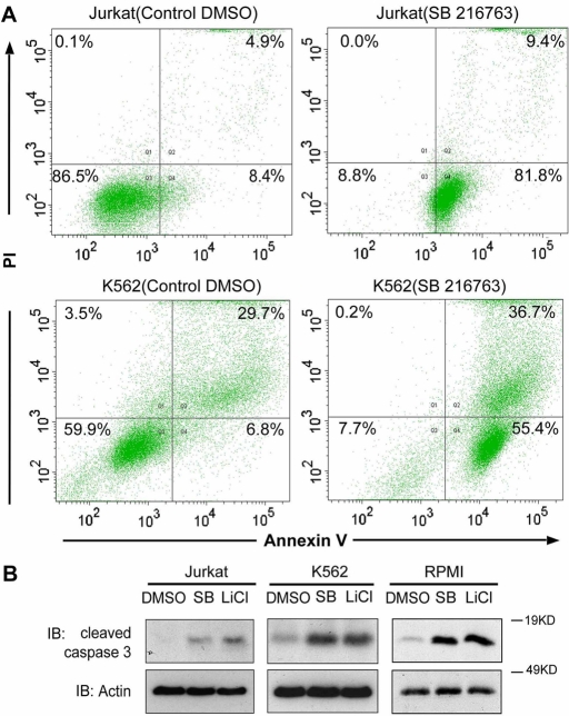
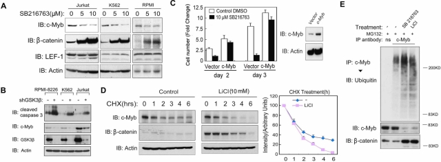
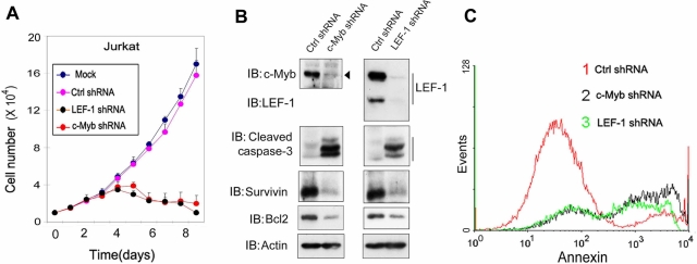
Glycogen synthase kinase 3β (GSK3β) regulates diverse physiological processes, including metabolism, development, oncogenesis, and neuroprotection. GSK3β kinase activity has been reported to be critical for various types of cancer cells, but the mechanism has remained elusive. In this study we examine the mechanism by which GSK3β regulates the survival of leukemia cells. We demonstrate that upon GSK3β kinase inhibition different types of leukemia cells show severe proliferation defects as a result of apoptosis. The transcription factor c-Myb is found to be the main target of GSK3β inhibition in cell survival. GSK3β inactivation reduces the expression of c-Myb by promoting its ubiquitination-mediated degradation, thereby inhibiting the expression of c-Myb-dependent antiapoptotic genes Bcl2 and survivin. Coimmunoprecipitation, reporter assays, chromatin immunoprecipitation, and knockdown studies show that c-Myb needs to interact and cooperate with transcription factor LEF-1 in the activation of Bcl2 and survivin and that both transcription factors are required for cell survival. These data reveal an as-yet-unknown mechanism by which GSK3β controls cell survival.


Figures

FIGURE 1:
FIGURE 1:
GSK3β inactivation causes growth inhibition of leukemia cells. (A) Jurkat, K562, RPMI 8226, and HEK293 cells were treated with or without LiCl (10 mM) or SB216763 (10 μM). Cell numbers were determined with a CASY model TT cell counter (Schärfe System, Reutlingen, Germany) on days 0 and 3; the fold change is depicted. (B) Cells were treated with or without SB216763 as described, and their viability was measured with the CellTiter 96 AQueous One Solution Cell Proliferation Assay (MTS; Promega, Madison, WI) at indicated time points. The relative viability (±SB216763) is depicted. (C) Jurkat cells were infected with lentivirus expressing GSK3β WT, GSK3β S9A, and GSK3β K85M and treated with or without SB216763 (10 μM). Cell numbers were determined as described for A. (D) Immunoblotting shows the expression levels of wild-type and mutant GSK3β in the experiment shown in C and verification of their effects on β-catenin expression. Data are represented as mean and SD of triplicates and are representative of at least two independent experiments.
FIGURE 2:
FIGURE 2:
GSK3β inactivation induces apoptosis in leukemia cells. (A) Jurkat and K562 cells were treated with or without SB216763 (10 μM) for 20 h, stained with annexin V and PI, and analyzed by fluorescence-activated cell sorting. (B) Jurkat, K562, and RPMI 8226 cells were treated with SB216763 (SB; 10 μM) or LiCl (10 mM) for 20 h, and cell lysates were analyzed by immunoblotting.
FIGURE 3:
FIGURE 3:
GSK3β inactivation inhibits the expression of Bcl2 and survivin. (A) Jurkat cells were treated with SB216763 (10 μM) or LiCl (10 mM) for 20 h, and RNA was reverse transcribed and analyzed by qRT-PCR. Values and error bars represent the mean and SD of triplicates and are representative of at least two independent experiments. (B) Jurkat, K562, and RPMI 8226 cells were treated with SB216763 (SB) (10 μM) or LiCl (10 mM) for 20 h, and cell lysates were analyzed by immunoblotting. (C, D) Jurkat cells were treated with or without SB216763 (10 μM) for 8 h. Cells were lysed, and ChIP was performed with anti–LEF-1, anti–c-Myb, or a control antibody (immunoglobulin G [IgG]). The precipitated promoter fragments of Bcl2 (C) and survivin (D) were amplified by qRT-PCR (top). The positions of the identified LEF-1– and c-Myb–binding sites in the promoters are shown in a sketch map (bottom). Values and error bars represent the mean and SD of triplicates and are representative of at least two independent experiments.
FIGURE 4:
FIGURE 4:
The c-Myb protein level is critical for GSK3β-dependent cell survival. (A) Jurkat, K562, and RPMI 8226 cells were treated with or without 5 or 10 μM SB216763 for 8 h, and cell lysates were analyzed by immunoblotting. (B) RPMI 8226, K562, and Jurkat cells were infected with lentivirus expressing GSK3β shRNA or a nonspecific control shRNA. Four days after infection, cells were harvested for immunoblotting analysis. (C) Jurkat cells were infected with lentiviral-overexpressed c-Myb or a control vector and after 48 h treated with or without SB216763 (10 μM) for another 2 or 3 d. Cell numbers were determined at days 0, 2, and 3 with a CASY Model TT cell counter (left). Data are represented as mean and SD. The overexpression of c-Myb was verified by immunoblotting (right). (D) Jurkat cells were treated with cycloheximide (20 μg/ml) in the absence or presence of LiCl (10 mM) for the indicated time points, and cell lysates were analyzed by immunoblotting. For the quantification on the right, the c-Myb protein level at time 0 was set at 100%. (E) Jurkat cells were treated with or without SB216763 (10 μM) or LiCl (10 mM) for 6 h, after which MG132 (10 μM) was added for another 4 h. c-Myb and nonspecific control immunoprecipitations were analyzed by SDS–PAGE and immunoblotting.
FIGURE 5:
FIGURE 5:
LEF-1 and c-Myb cooperatively regulate transcription. (A–D) K562 cells were transiently transfected with the indicated plasmids and analyzed for luciferase assay after 36 h. Data are represented as mean and SD. (E) Jurkat cells were infected with lentivirus expressing c-Myb or LEF-1 shRNA or a nonspecific control (ctrl) shRNA. After 60 h, cells were lysed and ChIP was performed with anti–LEF-1, anti–c-Myb, or a control antibody (IgG). The precipitated promoter fragments were amplified by qRT-PCR (left). Data are represented as mean and SD. The knockdown efficiency of the c-Myb and LEF-1 shRNAs was validated by immunoblotting analysis (right). (F) To detect the interaction between c-Myb and LEF-1 in Jurkat cells, LEF-1 and nonspecific control (ns) immunoprecipitations (IPs) were analyzed by SDS–PAGE and immunoblotting (IB). (G) RNA from the Jurkat cells treated as described for E was analyzed by qRT-PCR analysis. Values and error bars represent the mean and SD of triplicates and are representative of at least two independent experiments.
FIGURE 6:
FIGURE 6:
C-Myb and LEF-1 are both required for the survival of leukemia cells. (A) Jurkat cells were infected with lentivirus expressing c-Myb or LEF-1 shRNA, or nonspecific control (ctrl) shRNA or were mock infected. The cell numbers were counted at the indicated days. (B) Lysates of Jurkat cells treated as described for A were isolated 60 h after infection and analyzed by immunoblotting. (C) Jurkat cells treated as described for A were harvested 60 h after infection, stained with annexin V and PI, and analyzed by fluorescence-activated cell sorting.

References

    1. Baki L, Shioi J, Wen P, Shao Z, Schwarzman A, Gama-Sosa M, Neve R, Robakis NK. PS1 activates PI3K thus inhibiting GSK-3 activity and tau overphosphorylation: effects of FAD mutations. EMBO J. 2004;23:2586–2596. - PMC - PubMed
    1. Behrens J, von Kries JP, Kuhl M, Bruhn L, Wedlich D, Grosschedl R, Birchmeier W. Functional interaction of beta-catenin with the transcription factor LEF-1. Nature. 1996;382:638–642. - PubMed
    1. Beug H, von Kirchbach A, Doderlein G, Conscience JF, Graf T. Chicken hematopoietic cells transformed by seven strains of defective avian leukemia viruses display three distinct phenotypes of differentiation. Cell. 1979;18:375–390. - PubMed
    1. Clarke MF, Kukowska-Latallo JF, Westin E, Smith M, Prochownik EV. Constitutive expression of a c-myb cDNA blocks Friend murine erythroleukemia cell differentiation. Mol Cell Biol. 1988;8:884–892. - PMC - PubMed
    1. Corradini F, Cesi V, Bartella V, Pani E, Bussolari R, Candini O, Calabretta B. Enhanced proliferative potential of hematopoietic cells expressing degradation-resistant c-Myb mutants. J Biol Chem. 2005;280:30254–30262. - PubMed

Publication types

MeSH terms

LinkOut - more resources