. 2011 Nov;92(Pt 11):2485-2493.
doi: 10.1099/vir.0.036715-0. Epub 2011 Jul 27.

Ultracentrifugation deforms unfixed influenza A virions

Affiliations

Ultracentrifugation deforms unfixed influenza A virions

Yukihiko Sugita et al. J Gen Virol. 2011 Nov.

Abstract

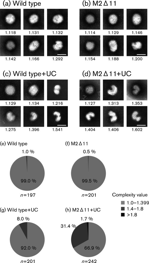
Negatively stained influenza virions sometimes show irregular morphology and are often referred to as pleomorphic. However, this irregular morphology has not been visualized when ultrathin-section transmission and scanning electron microscopies are used. This study focused on the effects of ultracentrifugation on influenza A virion morphology, as negative staining often involves ultracentrifugation to concentrate or purify virions. The morphologies of unfixed, glutaraldehyde-fixed and osmium tetroxide-fixed virions were quantitatively compared before and after ultracentrifugation, and it was found that, without chemical fixation, approximately 30% of virions were altered from oval to irregular shapes following ultracentrifugation. By contrast, most glutaraldehyde-fixed virions remained uniformly elliptical, even after ultracentrifugation. When a virus with an 11 aa deletion at the C terminus of its M2 cytoplasmic tail was ultracentrifuged, its morphology was appreciably deformed compared with that of the wild-type virus. These results demonstrate that the native morphology of influenza A virions is regular but is disrupted by ultracentrifugation, and that the cytoplasmic tail of M2 is important for virion integrity.


Figures

Fig. 1.
Fig. 1.
Budding PR8 virions observed by ultrathin-section TEM. (a) A representative electron micrograph of PR8 virions budding from the cells of allantoic membranes. Uniformly oval virions can be seen budding from the apical plasma membrane. (b) Representative electron micrographs of ultrathin-sectioned virions. The respective shape complexity values are given below the photos. (c) Proportion of shape complexity values of ultrathin-sectioned virions. In total, 216 virions were assessed. Bars, 500 nm (a); 100 nm (b).
Fig. 2.
Fig. 2.
PR8 virions observed by SEM. (a) A representative electron micrograph of PR8 virions budding from the cells of allantoic membranes. (b) A representative electron micrograph of virions adsorbed onto a chicken erythrocyte, which was found with allantoic membrane cells under an SEM field. Elliptical and spherical virions can be seen. (c) A higher magnification of the representative electron micrographs of released virions on chicken erythrocytes. Respective shape complexity values are given below the photos. (d) Proportion of shape complexity values of virions visualized by SEM. In total, 197 virions were examined. Bars, 500 nm (a, b); 100 nm (c).
Fig. 3.
Fig. 3.
Negatively stained PR8 virions. Representative electron micrographs of unfixed virions (a), OsO4-fixed virions (b), GLA-fixed virions (c), unfixed and ultracentrifuged (UC) virions (d), OsO4-fixed and UC virions (e) and GLA-fixed and UC virions (f). The respective shape complexity values are given below the photos. The proportion of shape complexity values for samples (a)–(f) is shown in (g)–(l), respectively. The total number of analysed virions is shown beneath the graphs. Bars, 100 nm.
Fig. 4.
Fig. 4.
Examples of irregular-shaped PR8 virions with complexity values >1.8. (a–c) Unfixed and non-ultracentrifuged virions; (d–g) unfixed and ultracentrifuged virions. Bars, 100 nm.
Fig. 5.
Fig. 5.
Comparison of WSN wild-type and M2Δ11 virions. (a, b) Western blot analysis of the structural proteins of the two viruses. (a) Viral proteins HA, NP and M1 were separated by SDS-PAGE under non-reducing conditions and detected with an anti-influenza virus rabbit polyclonal antibody (R309). (b) Under reducing conditions, monomeric M2 proteins were detected with an anti-M2 mAb (14C2). (c, d) WSN wild-type and M2Δ11 virions observed by ultrathin-section TEM. Rod-like RNPs can be seen in all of the virions. Bars, 100 nm.
Fig. 6.
Fig. 6.
Negatively stained WSN virions. Representative electron micrographs of WSN wild-type virions (a), M2Δ11 virions (b), ultracentrifuged (UC) wild-type virions (c) and UC M2Δ11 virions. The proportion of shape complexity values for samples (a)–(d) is shown in (e)–(h), respectively. Respective shape complexity values are given below the panels. The total number of analysed virions is shown beneath the graphs. Bars, 100 nm.

References

    1. Almeida J. D., Waterson A. P. (1967). Some observations on the envelope of an influenza virus. J Gen Microbiol 46, 107–110 - PubMed
    1. Almeida J. D., Waterson A. P., Drewe J. A. (1967). A morphological comparison of Bittner and influenza viruses. J Hyg (Lond) 65, 467–474 10.1017/S0022172400046003 - DOI - PMC - PubMed
    1. Bächi T., Gerhard W., Lindenmann J., Mühlethaler K. (1969). Morphogenesis of influenza A virus in Ehrlich ascites tumor cells as revealed by thin-sectioning and freeze-etching. J Virol 4, 769–776 - PMC - PubMed
    1. Booy F. P., Ruigrok R. W., van Bruggen E. F. (1985). Electron microscopy of influenza virus. A comparison of negatively stained and ice-embedded particles. J Mol Biol 184, 667–676 10.1016/0022-2836(85)90312-2 - DOI - PubMed
    1. Boulan E. R., Sabatini D. D. (1978). Asymmetric budding of viruses in epithelial monolayers: a model system for study of epithelial polarity. Proc Natl Acad Sci U S A 75, 5071–5075 10.1073/pnas.75.10.5071 - DOI - PMC - PubMed

Publication types