. 2011 Jul 27;31(30):10819-28.
doi: 10.1523/JNEUROSCI.1642-11.2011.

Peripheral nerve injury alters blood-spinal cord barrier functional and molecular integrity through a selective inflammatory pathway

Affiliations

Peripheral nerve injury alters blood-spinal cord barrier functional and molecular integrity through a selective inflammatory pathway

Stefania Echeverry et al. J Neurosci. .

Abstract

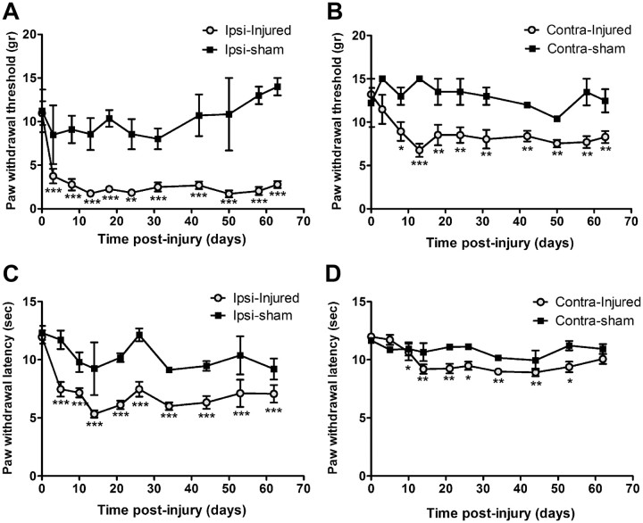
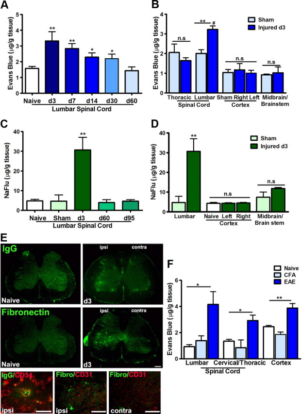
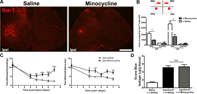
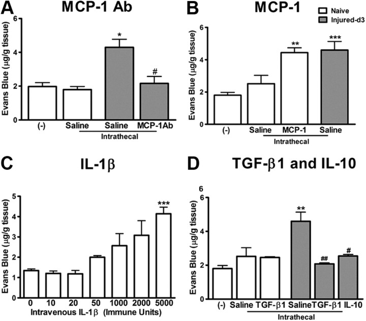
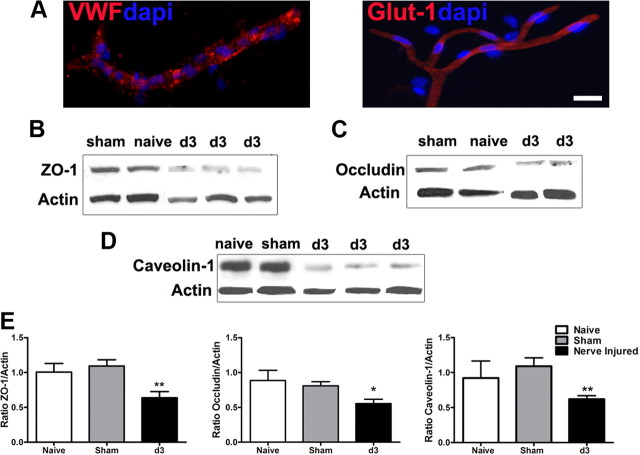
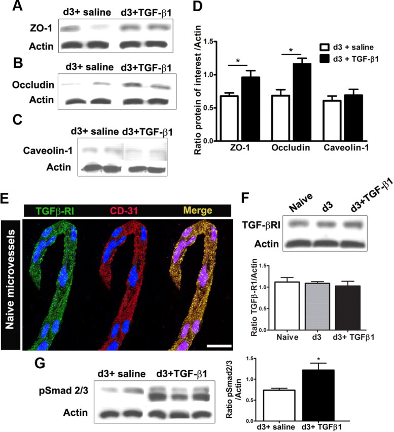
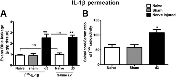
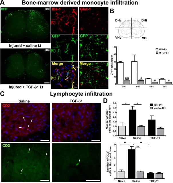
Peripheral nerve lesion triggers alterations in the spinal microenvironment that contribute to the pathogenesis of neuropathic pain. While neurons and glia have been implicated in these functional changes, it remains largely underexplored whether the blood-spinal cord barrier (BSCB) is also involved. The BSCB is an important component in the CNS homeostasis, and compromised BSCB has been associated with different pathologies affecting the spinal cord. Here, we demonstrated that a remote injury on the peripheral nerve in rats triggered a leakage of the BSCB, which was independent of spinal microglial activation. The increase of BSCB permeability to different size tracers, such as Evans Blue and sodium fluorescein, was restricted to the lumbar spinal cord and prominent for at least 4 weeks after injury. The spinal inflammatory reaction triggered by nerve injury was a key player in modulating BSCB permeability. We identified MCP-1 as an endogenous trigger for the BSCB leakage. BSCB permeability can also be impaired by circulating IL-1β. In contrast, antiinflammatory cytokines TGF-β1 and IL-10 were able to shut down the openings of the BSCB following nerve injury. Peripheral nerve injury caused a decrease in tight junction and caveolae-associated proteins. Interestingly, ZO-1 and occludin, but not caveolin-1, were rescued by TGF-β1. Furthermore, our data provide direct evidence that disrupted BSCB following nerve injury contributed to the influx of inflammatory mediators and the recruitment of spinal blood borne monocytes/macrophages, which played a major role in the development of neuropathic pain. These findings highlight the importance of inflammation in BSCB integrity and in spinal cord homeostasis.


Figures

Figure 1.
Figure 1.
Development of neuropathic pain following injury on sciatic nerve. Shortly after the partial ligation on the sciatic nerve, rats develop mechanical allodynia and thermal hyperalgesia in the ipsilateral hindpaws. Withdrawal threshold of both ipsilateral (A) and contralateral (B) paws to calibrated von Frey hair stimulation decreased significantly, starting from day 3 until at least day 63 after injury. Withdrawal latency to noxious heat stimuli was also decreased at ipsilateral side (C), and to a less extent at the contralateral paw (D). N = 6/group. Values are presented as means ± SEM. *p < 0.05, **p < 0.01, ***p < 0.001 from baseline.
Figure 2.
Figure 2.
Peripheral nerve injury impairs BSCB permeability. A, Evans Blue (MW, 961 Da; EB-albumin, MW, 68,500 Da) extravasation within lumbar spinal cords at different time points after nerve injury. Note there is a prominent increase of EB content within lumbar spinal cords during the first 4 weeks after nerve injury, which returns to normal levels at 2 months. B, Quantification of EB leakage in different regions of the spinal cord and brain at day 3 after injury; the increase of EB is only significant in the lumbar spinal cord. C, NaFlu (MW, 376 Da) content at different time points after nerve injury. At late time points (>2 months), while BSCB permeability to EB is back to normal levels, the passage of small molecular (NaFlu) through BSCB is at the similar levels of naive animals as well; however, the increase of NaFlu at day 3 after injury is dramatic. D, NaFlu content in lumbar and brain regions at day 3 after injury. A significant extravasation is only observed in the lumbar segment. E, Leakage of plasma protein IgG and fibronectin in the spinal parenchyma 3 d after nerve injury with preference in the ipsilateral side. Endothelium markers CD31 and CD34 delineated blood vessels and confirmed the presence of plasma protein in the parenchyma. F, Evans Blue extravasation is generalized throughout the spinal cord and brain in EAE mice. Values are presented as means ± SEM. **p < 0.01, *p < 0.05 versus naive or sham; #p < 0.05 versus thoracic segment. N = 6–10/group. Scale bar, 50 μm.
Figure 3.
Figure 3.
BSCB disruption is not affected by the suppression of spinal microglial activation. A, Photomicrographs depicting Iba-1 staining in the lumbar spinal cord. Partial sciatic nerve ligation induced a remarkable increase of Iba-1 IR at the ipsilateral side in the dorsal and ventral horns. Intrathecal minocycline (150 μg/d for 7 d) successfully reduced this Iba-1+ IR. B, Quantification of the area occupied by the Iba-1+ IR reveals a striking reduction of microglial activation in the minocycline-treated animals compared with saline-treated animals. C, Mechanical allodynia (left panel) and thermal hyperalgesia (right panel) were partially attenuated in minocycline-treated animals. D, Despite inhibition of microglia, EB leakage into the spinal cord remained elevated in minocycline-treated animals at day 7. Data are shown as means ± SEM. *p < 0.05, **p < 0.01, ***p < 0.001, minocycline-treated versus saline-treated groups. N = 6/group. Scale bar, 250 μm.
Figure 4.
Figure 4.
Inflammatory mediators modulate BSCB integrity. A, Neutralizing endogenous MCP-1 with a MCP-1 antibody (3 μg for 3 d) significantly reduced the leakage of EB in the spinal cord observed at day 3 after injury. B, Intrathecal infusion of recombinant murine MCP-1 (2.5 μg for 3 d) produced an increase of EB content in the spinal cord in naive animals, similar to that seen in nerve-injured animals. Intrathecal catheterization provoked a slight, but nonsignificant increase of EB content in the spinal cord. C, IL-1β injected intravenously caused a dose-dependent impairment of BSCB permeability to EB. D, Intrathecal administration of antiinflammatory cytokines TGF-β1 (2 μg) and IL-10 (3 μg) for 3 d significantly reduced the EB content in the spinal cord increased by peripheral nerve injury. TGF-β1 did not affect the BSCB permeability in naive rats. N = 3–6/group. Values are presented as means ± SEM. *p < 0.05, **p < 0.01, ***p < 0.001 versus naive rats without any treatment; #p < 0.05 versus injured saline-treated.
Figure 5.
Figure 5.
Downregulation of tight junction proteins following peripheral nerve injury. Microvessels isolated from the spinal cord were positive for endothelium markers, VWF and Glut-1 (A). The expression of tight junction-associated protein ZO-1 (B), occludin-1 (C), and caveolin-1, a major player in the formation of caveolae (D), was significantly lower in microvessels of animals day 3 after nerve lesion than controls (naive and sham). Quantitative assessment of protein levels is illustrated in E. Three separate experiments in which each treatment group consists of pooled microvessels from two animals were included. Values are presented as means ± SEM. *p < 0.05, **p < 0.01. Scale bar, 20 μm.
Figure 6.
Figure 6.
TGF-β1 alters tight junction protein levels in spinal cord microvessels. Intrathecal administration of TGF-β1 (2.5 μg for day 0 to day 3) successfully prevents the decrease in tight junction protein levels ZO-1 (A, D) and occludin (B, D) observed after peripheral nerve injury. Levels of caveolae structural component caveolin-1, however, remained unchanged (C, D). The receptor for TGF-β1 (TGF-β-RI) is found in isolated spinal cord endothelial cells, confirmed by the colocalization with vascular marker CD31. DAPI staining (blue) was used for cellular identification (E). The protein levels of the TGF-β-RI remain unchanged after peripheral nerve injury, with or without TGF-β1 infusion (F). TGF-β1 treatment induced the phosphorylation of signaling pathway proteins Smad2/3 (pSmad2/3) in spinal cord microvessels (G). Three separate experiments in which each treatment group consists of pooled microvessels from two animals were included. Values are presented as means ± SEM. *p < 0.05 versus d3 + saline. Scale bar, 10 μm.
Figure 7.
Figure 7.
Peripheral nerve injury induced disruption of BSCB provided access to circulating immune mediators into the spinal cord parenchyma. A, Intracardiac injection of 125I-IL-1β (0.7 μCi/150 μl) did not affect BSCB permeability to EB in either naive or nerve-injured rats. B, There was a significant increase in the uptake of 125I-IL-1β in the lumbar spinal cord 3 d after sciatic nerve injury compared with control groups. Values are presented as means ± SEM. *p < 0.05, **p < 0.01 versus naive.
Figure 8.
Figure 8.
Peripheral nerve injury-induced disruption of BSCB provided access to circulating immune cells into the spinal cord parenchyma. A, GFP+ bone marrow-derived cells infiltrated into the spinal cords after PNI, which differentiated into Iba-1+ microglia. Intrathecal injection of TGF-β1 prevented the entrance of circulating immune cells at 14 d after nerve injury. Evidence on localization of infiltrated GFP+ bone marrow-derived cells in the parenchyma is shown by coimmunostaining with vessel marker Glut-1. Scale bars: left, 250 μm; middle, 10 μm; right, 20 μm. B, Quantitative analysis of GFP+ cells in the spinal cords of GFP chimeric mice after PNI and TGF-β1 treatment. C, To compare with naive animals, there was a marked infiltration of CD2+ and CD3+ lymphocytes into the ipsilateral side of spinal cords after peripheral nerve injury (white arrows). Intrathecal injection of TGF-β1 prevented the entrance of circulating lymphocytes at 7 d after nerve injury. Scale bar, 50 μm. D, Quantitative analysis of CD2+ and CD3+ cells in the dorsal horns of spinal cords of rats after nerve injury and TGF-β1 treatment. TGF-β1 successfully reduced the number of cells invading the spinal parenchyma. N = 6/group. Values are expressed as means ± SEM. *p < 0.05, **p < 0.01 versus controls (naive and sham); #p < 0.05, ##p < 0.01, ###p < 0.001 versus saline.

References

    1. Andjelkovic AV, Spencer DD, Pachter JS. Visualization of chemokine binding sites on human brain microvessels. J Cell Biol. 1999;145:403–412. - PMC - PubMed
    1. Banks WA, Erickson MA. The blood-brain barrier and immune function and dysfunction. Neurobiol Dis. 2010;37:26–32. - PubMed
    1. Basbaum AI, Bautista DM, Scherrer G, Julius D. Cellular and molecular mechanisms of pain. Cell. 2009;139:267–284. - PMC - PubMed
    1. Beggs S, Liu XJ, Kwan C, Salter MW. Peripheral nerve injury and TRPV1-expressing primary afferent C-fibers cause opening of the blood-brain barrier. Mol Pain. 2010;6:74. - PMC - PubMed
    1. Bennett J, Basivireddy J, Kollar A, Biron KE, Reickmann P, Jefferies WA, McQuaid S. Blood-brain barrier disruption and enhanced vascular permeability in the multiple sclerosis model EAE. J Neuroimmunol. 2010;229:180–191. - PubMed

Publication types

MeSH terms

Substances

LinkOut - more resources