. 2011 Jul 27;31(30):10859-71.
doi: 10.1523/JNEUROSCI.0445-11.2011.

In vivo neuronal subtype-specific targets of Atoh1 (Math1) in dorsal spinal cord

Affiliations

In vivo neuronal subtype-specific targets of Atoh1 (Math1) in dorsal spinal cord

Helen C Lai et al. J Neurosci. .

Abstract

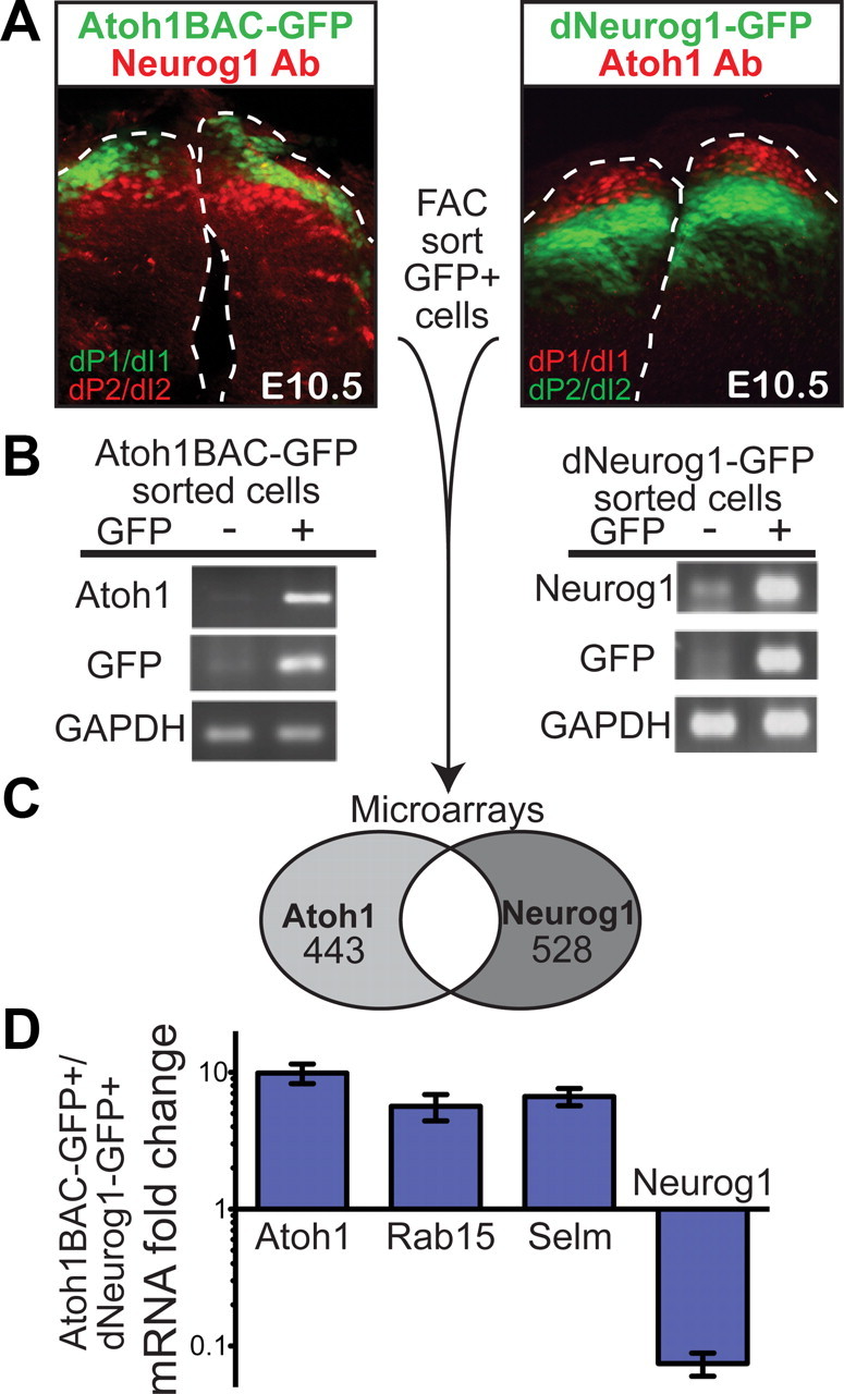
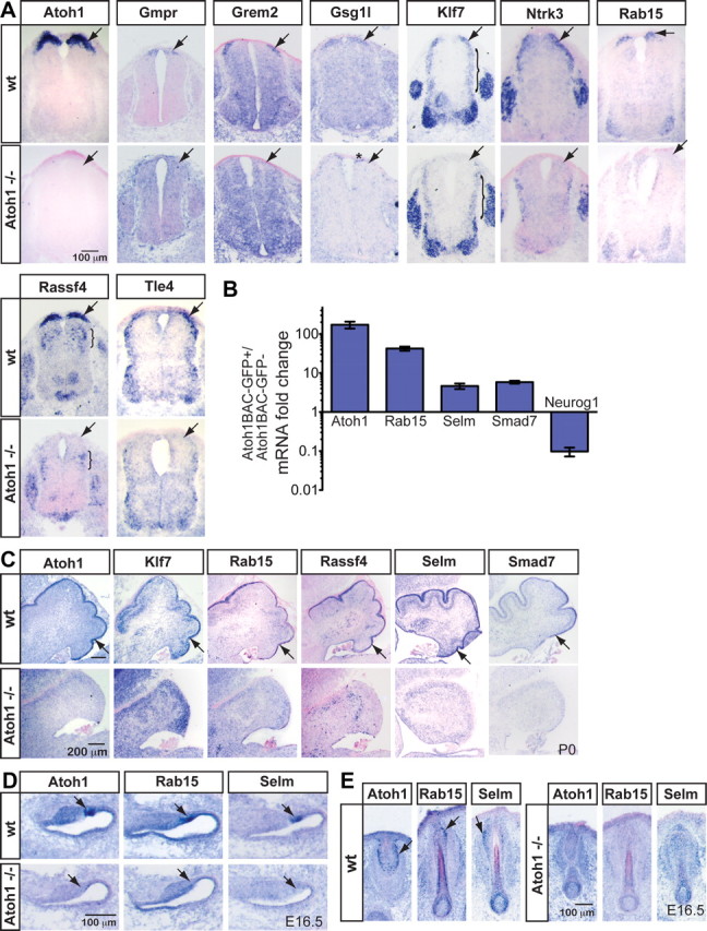
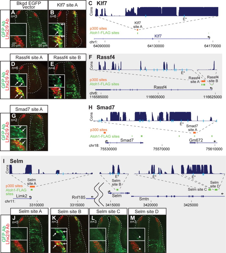
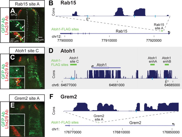
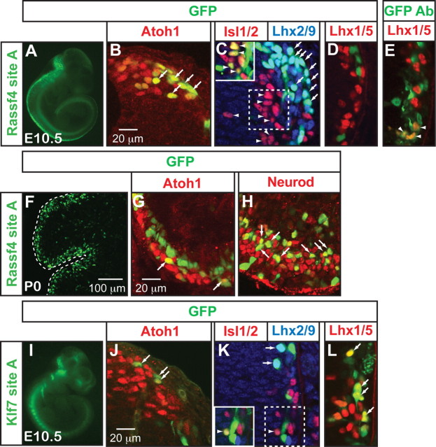
Neural basic helix-loop-helix (bHLH) transcription factors are crucial in regulating the differentiation and neuronal subtype specification of neurons. Precisely how these transcription factors direct such processes is largely unknown due to the lack of bona fide targets in vivo. Genetic evidence suggests that bHLH factors have shared targets in their common differentiation role, but unique targets with respect to their distinct roles in neuronal subtype specification. However, whether neuronal subtype-specific targets exist remains an unsolved question. To address this question, we focused on Atoh1 (Math1), a bHLH transcription factor that specifies distinct neuronal subtypes of the proprioceptive pathway in mammals including the dI1 (dorsal interneuron 1) population of the developing spinal cord. We identified transcripts unique to the Atoh1-derived lineage using microarray analyses of specific bHLH-sorted populations from mouse. Chromatin immunoprecipitation-sequencing experiments followed by enhancer reporter analyses identified five direct neuronal subtype-specific targets of Atoh1 in vivo along with their Atoh1-responsive enhancers. These targets, Klf7, Rab15, Rassf4, Selm, and Smad7, have diverse functions that range from transcription factors to regulators of endocytosis and signaling pathways. Only Rab15 and Selm are expressed across several different Atoh1-specified neuronal subtypes including external granule cells (external granule cell layer) in the developing cerebellum, hair cells of the inner ear, and Merkel cells. Our work establishes on a molecular level that neuronal differentiation bHLH transcription factors have distinct lineage-specific targets.


Figures

Figure 1.
Figure 1.
Discrete Atoh1 and Neurog1 dorsal neural tube populations were isolated for microarray analyses. A, Immunostaining of E10.5 mouse neural tubes (transverse sections) from Atoh1BAC-GFP and dNeurog1-GFP transgenic mice shows that the GFP fluorescence has little overlap with the neighboring population (immunostained with Neurog1 antibody or Atoh1 antibody, respectively). Atoh1 and Atoh1BAC-GFP mark the dorsal progenitor and interneuron 1 (dP1 and dI1) populations, while Neurog1 and dNeurog1-GFP mark the dorsal progenitor and interneuron 2 (dP2 and dI2) populations. GFP positive (+) and negative (−) cells were separated by FACS. B, RT-PCR of RNA extracted from GFP+ and GFP− cells shows that the cells were well separated with Atoh1 and GFP transcripts enriched in the GFP+ Atoh1BAC-GFP sorted cells, and Neurog1 and GFP transcripts enriched in the GFP+ dNeurog1-GFP sorted cells, while the control, GAPDH, remained in both populations. C, GFP+ cells from Atoh1BAC-GFP and dNeurog1-GFP neural tubes were analyzed by Affymetrix Mouse 430 2.0 microarrays and the number of genes enriched in each population is given. D, mRNA fold change expression of transcripts from Atoh1BAC-GFP+ sorted cells compared with dNeurog1-GFP+ cells quantitated by RT-qPCR. Ppib (formerly Cyclophilin B) was used as the endogenous control gene. SDs are reported.
Figure 2.
Figure 2.
Novel transcripts specific to Atoh1-derived domains were identified. A, Several transcripts found to be enriched in the Atoh1-derived population over the neighboring Neurog1-derived population in the microarray analyses are expressed in the dorsal-most part of the developing E10.5 neural tube and are absent in Atoh1 mutant embryos (arrows) (Ben-Arie et al., 1997) as detected by ISH. The asterisk (*) denotes that Gsg1l expression appears to be near the roof plate in the Atoh1 knock-out. The bracket (}) indicates lateral expression of Klf7 and expression of Rassf4 in the Ascl1-derived domain of the neural tube in their respective panels. B, mRNA fold change expression of transcripts from Atoh1BAC-GFP+ sorted cells compared with GFP− cells quantitated by RT-qPCR. Ppib (formerly Cyclophilin B) was used as the endogenous control gene. SDs are reported. C, Klf7, Rab15, Rassf4, Selm, and Smad7 are present in the developing EGL at P0. D, E, Rab15 and Selm are present in developing E16.5 hair cells (D) and Merkel cells in the vibrissae (E), and their expression is lost in the Atoh1 mutant mouse.
Figure 3.
Figure 3.
Identification of multiple enhancers of Atoh1 target genes that direct expression to the dI1 population by Atoh1-FLAG ChIP-seq. ChIP of Atoh1-FLAG (green), a FLAG-tagged Atoh1 knock-in mouse, from P5 cerebella, identified several binding regions near Atoh1 downstream genes (C, F, H, I). Regions bound by p300 (orange), a transcriptional coactivator, from E11.5 neural tubes are also noted. Gene structures are shown in blue and conservation across 30 different vertebrate species for the tested sites is shown in purple (UCSC browser) (Kent et al., 2002). E-boxes matching the consensus CANNTG (light blue lines) and the identified common E-box sequence, AMCAGMTG (E*), are shown. Genomic sites were tested for enhancer activity in GFP reporter assays in the chick neural tube with the right side being the electroporated side. GFP expression is labeled with GFP antibody (green) and the dI1 neurons labeled with Lhx2/9 antibody (red) (A, B, D, E, G, J–M). The insets highlight the dI1 domain [top, GFP antibody staining (green); bottom, GFP (green) and Lhx2/9 (red) antibody staining]. The arrows indicate double-labeled cells (B, D, G, K), and the arrowheads indicate low level of double labeling (E, M). The EGFP vector with no enhancer sequence (A) gives some background fluorescence in the ventral neural tube that does not overlap with the dI1 domain (A, inset).
Figure 4.
Figure 4.
Atoh1-FLAG bound sites were tested for enhancer activity. A, B, Rab15 site A in an intron of Rab15 directs expression to the dI1 domain in GFP reporter assays in the chick neural tube as detected by GFP antibody staining (green) colocalized with Lhx2/9 antibody staining (red) (arrows). C, D, Atoh1 site C (∼1 kb upstream of Atoh1) driving GFP gives partial expression in the dI1 domain (arrowhead), but not significantly. This is consistent with the previous report by our laboratory that a transgenic line of 15 kb sequence 5′ of the Atoh1 gene does not drive LacZ reporter expression in mice (Helms et al., 2000). E, F, Grem2 had only one Atoh1-FLAG peak within 200 kb of the gene in the Grem2 intron. Grem2 site A did not appear to drive GFP to the dI1 domain. Atoh1-FLAG sites (green), gene structure (blue), and vertebrate conservation of 30 species (purple) are displayed from UCSC browser (Kent et al., 2002). E-boxes matching the consensus CANNTG (light blue lines) and the newly identified AMCAGMTG (E*) common E-box are shown. The insets highlight the dI1 domain [top, GFP antibody staining (green); bottom, GFP (green) and Lhx2/9 (red) antibody staining].
Figure 5.
Figure 5.
Enhancers of Atoh1 targets are induced by Atoh1 and contain a common E-box motif. Each enhancer found to drive expression to dI1 neurons was tested for its sensitivity to Atoh1 overexpression in chick neural tubes. A–F″, Enhancers of Atoh1 targets, Klf7 site A (B–B″), Rassf4 site A (C–C″), Selm site B (D–D″), Smad7 site A (E–E″), and Rab15 site A (F–F″) driving GFP were coinjected with a myc-tagged control bHLH inactive mutant (Nakada et al., 2004a), a BOSS-tagged Atoh1 (Helms et al., 2000), or a myc-tagged Ascl1 (Nakada et al., 2004a). In all cases, Atoh1 increased the GFP fluorescence intensity of the enhancer reporter (B′, C′, D′, E′, F′), while Ascl1 did not except for Rassf4 site A (C″) and Rab15 site A (F″). The insets are enlarged manipulated images showing that the GFP fluorescence could be detected and the cells were injected (myc or BOSS antibody, red). For D, D″, and F, the GFP gain was set very low to accommodate the significant increase in GFP fluorescence upon coelectroporation with Atoh1, so there appears to be no GFP fluorescence; however, if the gain is increased, there is detectable GFP fluorescence (our observation) as seen in Figure 3K and 4A. The EGFP vector with no enhancer was not significantly induced by Atoh1 or Ascl1 (A–A″). G, Colocalized cells (GFP+ and myc+ or BOSS+) were outlined and the average pixel intensity per cell calculated for each image. Number of images quantified per sample are given in A–F″. All five enhancers were induced by Atoh1, whereas Selm site A, which does not direct expression to the dI1 domain, was not induced by Atoh1 (n = 3) over control (n = 2). H, MEME enhancer analysis of Atoh1-bound regions of Atoh1 responding enhancers identified a common E-box motif. I, Comparison of the Atoh1 common E-box motif to consensus E-box sequences derived for Atonal (Powell et al., 2004), Atoh1 in the cerebellum (AtEAM) (Klisch et al., 2011), Neurog/Neurod1 (Seo et al., 2007), Ascl1 (Castro et al., 2006), and MyoD (Cao et al., 2010). The arrowhead points to the first N site of the E-box that is different between the Atoh1 common E-box and Atonal consensus binding site. B = C/G/T, D = A/G/T, K = G/T, M = A/C, R = G/A, S = G/C, W= A/T, Y = C/T. J, The average pixel intensity per cell was calculated (as in G) for Klf7 site A with either one or both of the two E-boxes mutated (Emut 1, E*mut 2, and Emut 1&2) with and without Atoh1. E*mut indicates the E-box that meets the common E-box motif found in H. K, Similarly, the average pixel intensity per cell was calculated for Rassf4 site A with only the common E-box mutated (E*mut) with and without Atoh1. The number of images quantified per condition is given. For G, J, and K, SEMs are reported. *p < 0.05, **p < 0.001, ***p < 0.0001; n.s., not significant.
Figure 6.
Figure 6.
Enhancers Rassf4 site A and Klf7 site A drive GFP expression to Atoh1-derived domains in transgenic mice. A–E, Rassf4 site A drives GFP expression preferentially to the Atoh1-derived domains of the developing neural tube. Endogenous GFP fluorescence colocalizes with Atoh1+ (B) and Lhx2/9+ (C) cells (arrows). Very light GFP fluorescence colocalizes with Islet1/2+ cells (arrowheads; C, inset; gain increased to visualize colocalization), a marker for dI3 cells. Colocalization of Lhx1/5, a marker for dI2 cells, can only be visualized by adding GFP antibody to increase the GFP signal (compare D, E, arrowheads). F–H, Rassf4 site A drives GFP expression to the external granule cell layer of the developing cerebellum at P0. Endogenous GFP colocalizes with Atoh1 antibody staining (G, arrows) and the differentiating granule cells marked by Neurod antibody (H, arrows). I–L, Klf7 site A drives GFP expression to the dI1 domain as well as dI2 and dI3. Endogenous GFP fluorescence colocalizes with Atoh1+ (J, arrows), Lhx2/9+ cells (K, arrows), Lhx1/5+ (L, arrows), and barely with Islet1/2+ cells (K, arrowhead; inset; gain increased to visualized colocalization).

References

    1. Abramoff MD, Magelhaes PJ, Ram SJ. Image processing with image. J Biophotonics Int. 2004;11:36–42.
    1. Aerts S, Quan XJ, Claeys A, Naval Sanchez M, Tate P, Yan J, Hassan BA. Robust target gene discovery through transcriptome perturbations and genome-wide enhancer predictions in Drosophila uncovers a regulatory basis for sensory specification. PLoS Biol. 2010;8:e1000435. - PMC - PubMed
    1. Bailey TL, Elkan C. Fitting a mixture model by expectation maximization to discover motifs in biopolymers. Proc Int Conf Intell Syst Mol Biol. 1994;2:28–36. - PubMed
    1. Ben-Arie N, McCall AE, Berkman S, Eichele G, Bellen HJ, Zoghbi HY. Evolutionary conservation of sequence and expression of the bHLH protein Atonal suggests a conserved role in neurogenesis. Hum Mol Genet. 1996;5:1207–1216. - PubMed
    1. Ben-Arie N, Bellen HJ, Armstrong DL, McCall AE, Gordadze PR, Guo Q, Matzuk MM, Zoghbi HY. Math1 is essential for genesis of cerebellar granule neurons. Nature. 1997;390:169–172. - PubMed

Publication types

MeSH terms