. 2011 Jul 27;31(30):10937-47.
doi: 10.1523/JNEUROSCI.5302-10.2011.

Longitudinal development of human brain wiring continues from childhood into adulthood

Affiliations

Longitudinal development of human brain wiring continues from childhood into adulthood

Catherine Lebel et al. J Neurosci. .

Abstract

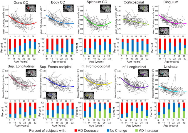
Healthy human brain development is a complex process that continues during childhood and adolescence, as demonstrated by many cross-sectional and several longitudinal studies. However, whether these changes end in adolescence is not clear. We examined longitudinal white matter maturation using diffusion tensor tractography in 103 healthy subjects aged 5-32 years; each volunteer was scanned at least twice, with 221 total scans. Fractional anisotropy (FA) and mean diffusivity (MD), parameters indicative of factors including myelination and axon density, were assessed in 10 major white matter tracts. All tracts showed significant nonlinear development trajectories for FA and MD. Significant within-subject changes occurred in the vast majority of children and early adolescents, and these changes were mostly complete by late adolescence for projection and commissural tracts. However, association tracts demonstrated postadolescent within-subject maturation of both FA and MD. Diffusion parameter changes were due primarily to decreasing perpendicular diffusivity, although increasing parallel diffusivity contributed to the prolonged increases of FA in association tracts. Volume increased significantly with age for most tracts, and longitudinal measures also demonstrated postadolescent volume increases in several association tracts. As volume increases were not directly associated with either elevated FA or reduced MD between scans, the observed diffusion parameter changes likely reflect microstructural maturation of brain white matter tracts rather than just gross anatomy.


Figures

Figure 1.
Figure 1.
Age at scans for all subjects. Each of the 221 scans obtained is represented by a circle; each of the 103 subjects is shown in a different row, with their scans connected by a straight line. Females (red) and males (blue) are marked separately. Note the relatively even spacing of subjects across the age range, with most subjects receiving two scans ∼4 years apart. Several subjects received three or four scans in total.
Figure 2.
Figure 2.
Longitudinal volume changes on T1-weighted images. Changes of white matter, gray matter, and total brain volume with age for each scan are shown (top row) along with bar graphs reflecting the percentage of subjects with volume increases (green), decreases (red), or no change (blue) within six age categories. White matter volume increased significantly across the age range, including the twenties, while gray matter volume decreased in the majority of children, adolescents, and young adults up to 25 years. These white matter increases and gray matter decreases offset one another such that total brain volume did not change in most persons, although many of the younger subjects had small increases.
Figure 3.
Figure 3.
Longitudinal age-related changes of fractional anisotropy. Individual FA data from 221 scans and bar graphs depicting the percentage of 103 subjects whose FA increased (green), decreased (red), or did not change (blue) in six age groupings are shown for all 10 white matter fibers. All fibers showed significant nonlinear increases of FA with age. In callosal and projection fibers, children and young adolescents had FA increases, while most young adults showed no change of FA (although 30% had increased FA of the body and splenium in 22–32 year group). The majority of young subjects also had FA increases in association fibers, yet importantly these fibers, such as the superior and inferior fronto-occipital, inferior longitudinal, and cingulum, continue to have 30–50% of subjects with FA increases between scans even in the 19–25 and 22–32 year age groups. Tracts displayed are from a 22-year-old male.
Figure 4.
Figure 4.
Longitudinal age-related changes of mean diffusivity. All 10 tracts had significant age-related decreases of MD. Commissural tracts demonstrated expected patterns, where a large proportion of subjects had decreasing MD in childhood and there were fewer subjects with decreases at older ages. Most association tracts, however, demonstrated more prolonged MD decreases, with 30–40% of older subjects having decreases of MD between scans. Notably, the inferior fronto-occipital, inferior longitudinal, and uncinate fasciculi show 40–60% of subjects with increasing MD between scans as early as the 11–19 year group. Other frontal lobe connecting white matter fibers, such as the genu of the corpus callosum and the cingulum, demonstrate 30–40% of subjects with increasing MD but only in the oldest age grouping of 22–32 years suggesting age-related decline is beginning in some subjects.
Figure 5.
Figure 5.
Sample tracts at two time points. Tracts are shown at two time points for several individuals. The superior longitudinal fasciculus (orange), corticospinal tract (green), and inferior longitudinal fasciculus (purple) are given as examples because they can be some of the more difficult and inconsistent fibers to track. Note that, although there are considerable variations in length, size, and shape of the tracts among individuals, the tracts look relatively similar within the same individual.
Figure 6.
Figure 6.
Longitudinal changes of parallel and perpendicular diffusivity. For all tracts, most young subjects had decreased perpendicular diffusivity between scans and fewer older subjects had decreases. Parallel diffusivity trajectories varied more among tracts. Most association fibers (such as the cingulum shown above) had only small changes of parallel diffusivity, although for tracts with prolonged FA changes (superior and inferior longitudinal, shown above, and inferior fronto-occipital fasciculi), a substantial portion of older subjects demonstrated increased parallel diffusivity between scans. Commissural fibers had substantial decreases of both parallel and perpendicular diffusivity in young subjects, with most subjects having no change at older ages. The corticospinal tract was unique in that its parallel diffusivity trajectory increased at young ages; its perpendicular diffusivity trajectory was similar to that of other tracts.
Figure 7.
Figure 7.
Longitudinal changes of tract volume. Volume changes were significant for most tracts, although not for the superior fronto-occipital fasciculus (no trend line shown). Most tracts had linear volume increases across the age range, although the cingulum, genu of the corpus callosum, and the inferior longitudinal fasciculus had quadratic trends with increases then decreases, and the splenium of the corpus callosum showed linear decreases of volume. Note that within-subject increases of tract volumes were observed in 40–50% of subjects in the 19–25 and 22–32 year age groups for the body of the corpus callosum, cingulum, and inferior longitudinal fasciculus.
Figure 8.
Figure 8.
Tract volume relationship with FA and MD changes. The percentage of subjects that showed increases (green), decreases (red), or no change (blue) in tract volume between scans are shown for each group depending on the evolution of FA or MD. While there is a decent proportion of subjects with elevated FA or reduced MD that have increased tract volume, there is a greater proportion of subjects with no change or even a decrease in tract volume in these cases. There is no clear association between tract volume and diffusion parameter changes.

Similar articles

Cited by

References

    1. Ashtari M, Cervellione KL, Hasan KM, Wu J, McIlree C, Kester H, Ardekani BA, Roofeh D, Szeszko PR, Kumra S. White matter development during late adolescence in healthy males: a cross-sectional diffusion tensor imaging study. Neuroimage. 2007;35:501–510. - PubMed
    1. Barnea-Goraly N, Menon V, Eckert M, Tamm L, Bammer R, Karchemskiy A, Dant CC, Reiss AL. White matter development during childhood and adolescence: a cross-sectional diffusion tensor imaging study. Cereb Cortex. 2005;15:1848–1854. - PubMed
    1. Bava S, Thayer R, Jacobus J, Ward M, Jernigan TL, Tapert SF. Longitudinal characterization of white matter maturation during adolescence. Brain Res. 2010;1327:38–46. - PMC - PubMed
    1. Beaulieu C. The basis of anisotropic water diffusion in the nervous system—a technical review. NMR Biomed. 2002;15:435–455. - PubMed
    1. Ben Bashat D, Ben Sira L, Graif M, Pianka P, Hendler T, Cohen Y, Assaf Y. Normal white matter development from infancy to adulthood: comparing diffusion tensor and high b value diffusion weighted MR images. J Magn Reson Imaging. 2005;21:503–511. - PubMed

Publication types