. 2011 Sep 1;187(5):2453-63.
doi: 10.4049/jimmunol.1101268. Epub 2011 Jul 27.

TCRs used in cancer gene therapy cross-react with MART-1/Melan-A tumor antigens via distinct mechanisms

Affiliations

TCRs used in cancer gene therapy cross-react with MART-1/Melan-A tumor antigens via distinct mechanisms

Oleg Y Borbulevych et al. J Immunol. .

Abstract

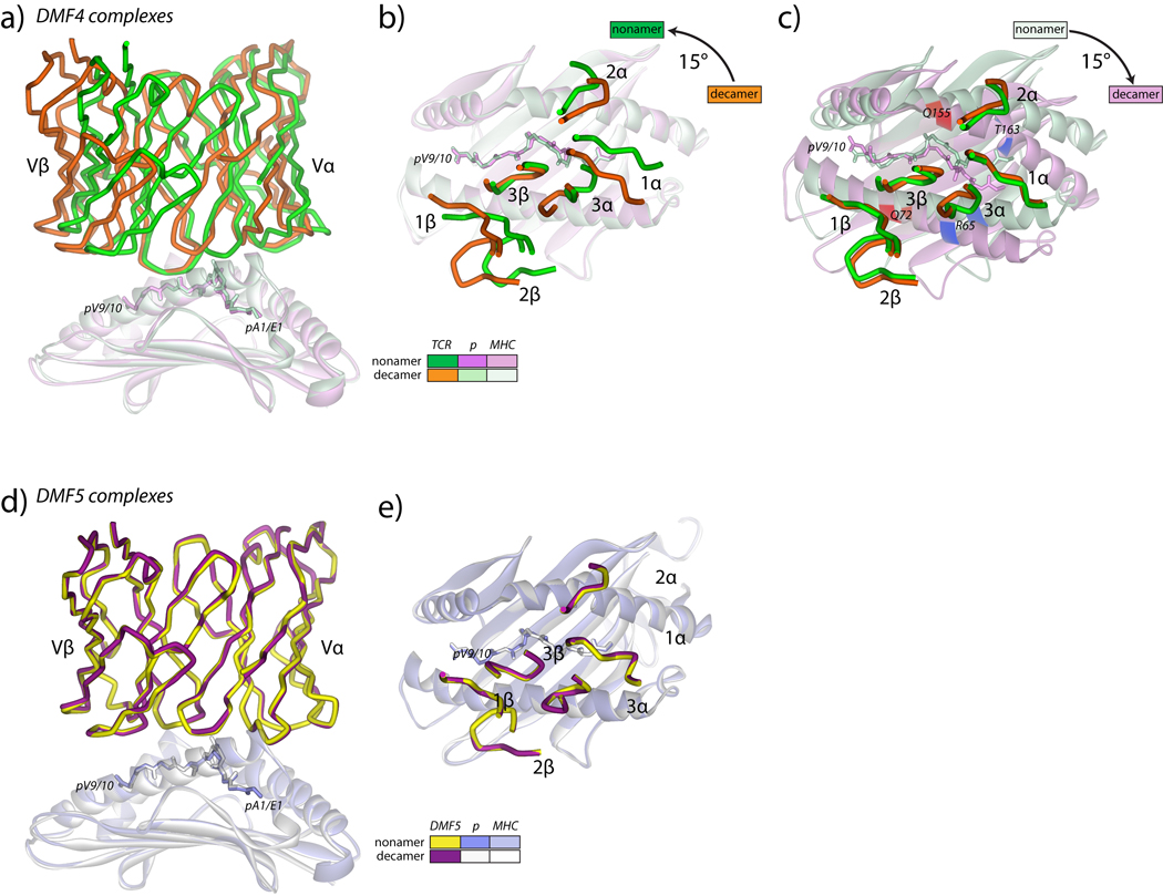
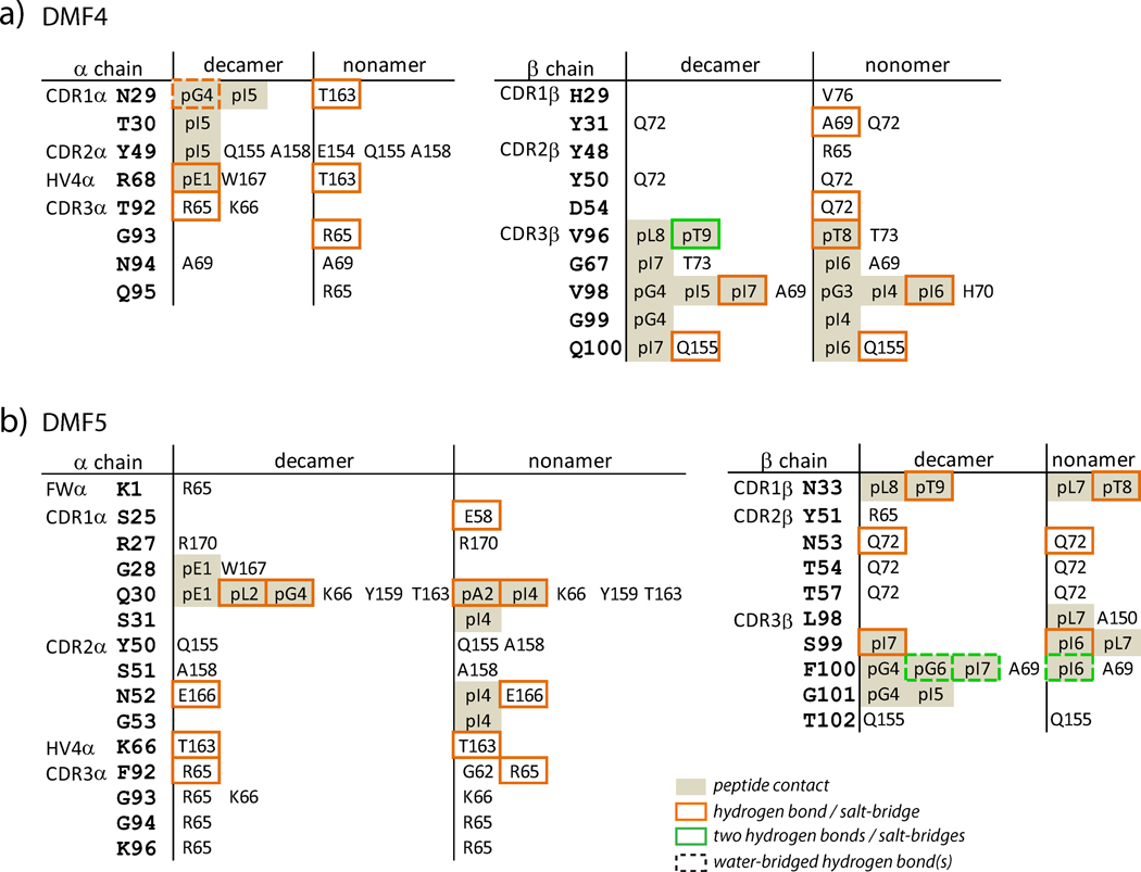
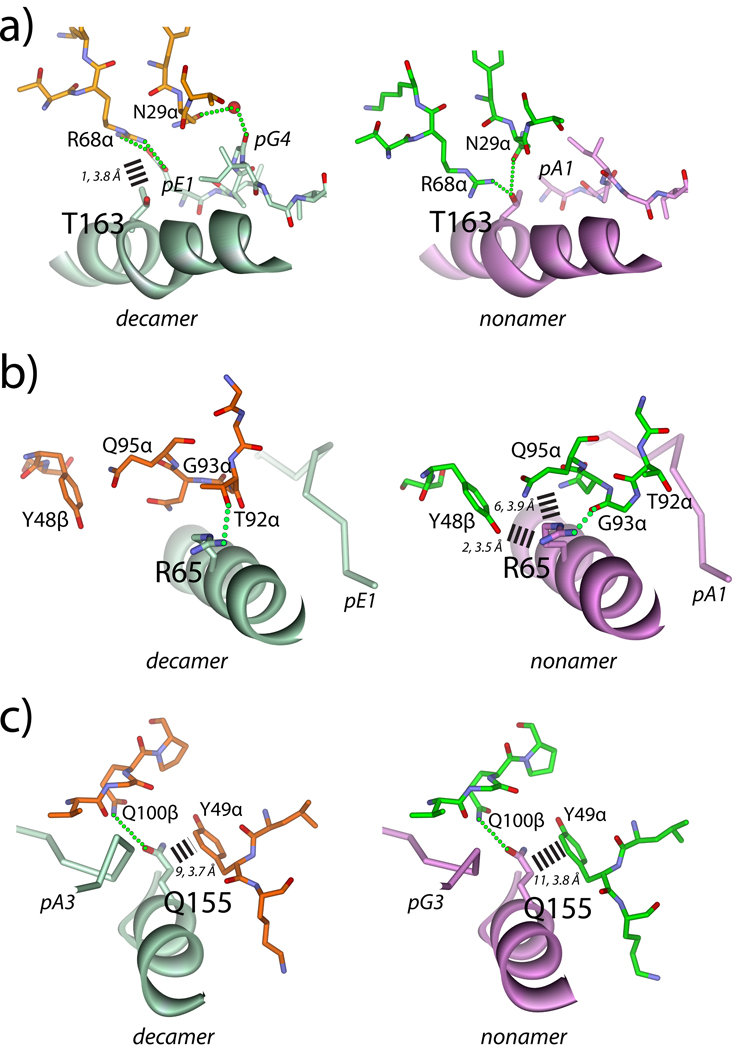
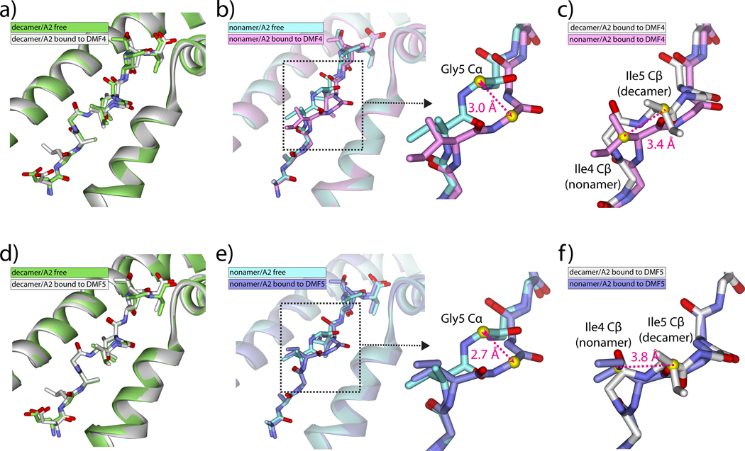
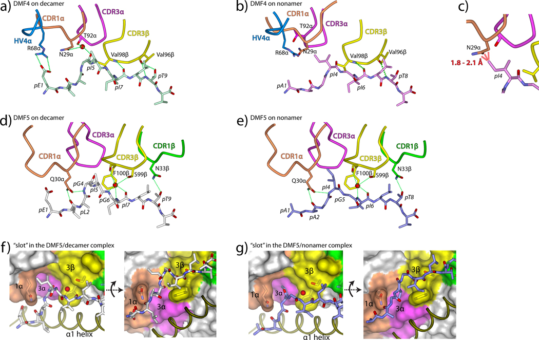
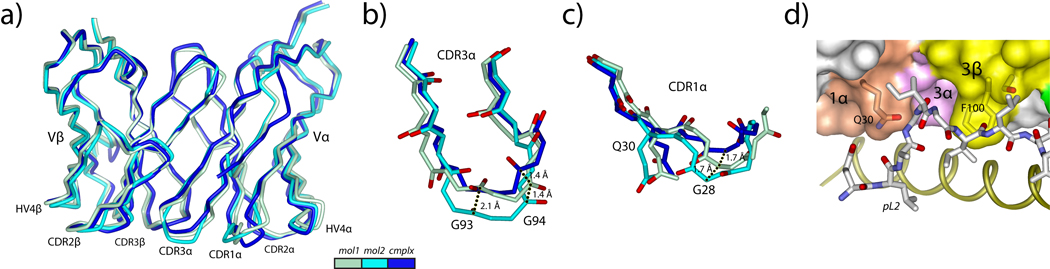
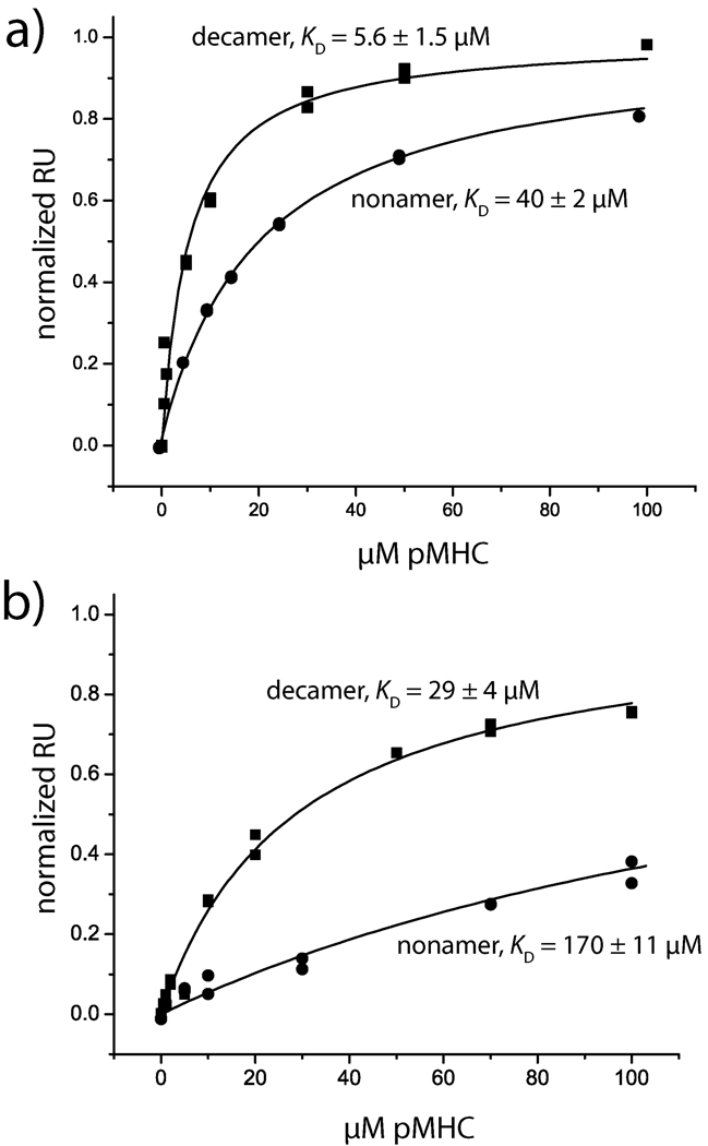
T cells engineered to express TCRs specific for tumor Ags can drive cancer regression. The first TCRs used in cancer gene therapy, DMF4 and DMF5, recognize two structurally distinct peptide epitopes of the melanoma-associated MART-1/Melan-A protein, both presented by the class I MHC protein HLA-A*0201. To help understand the mechanisms of TCR cross-reactivity and provide a foundation for the further development of immunotherapy, we determined the crystallographic structures of DMF4 and DMF5 in complex with both of the MART-1/Melan-A epitopes. The two TCRs use different mechanisms to accommodate the two ligands. Although DMF4 binds the two with a different orientation, altering its position over the peptide/MHC, DMF5 binds them both identically. The simpler mode of cross-reactivity by DMF5 is associated with higher affinity toward both ligands, consistent with the superior functional avidity of DMF5. More generally, the observation of two diverging mechanisms of cross-reactivity with the same Ags and the finding that TCR-binding orientation can be determined by peptide alone extend our understanding of the mechanisms underlying TCR cross-reactivity.


Figures

Figure 1
Figure 1
Overview of the DMF4 and DMF5 MART-1 nonamer and decamer peptide/HLA-A2 ternary complexes. A) Side view of the two DMF4 complexes, showing the differences in the TCR variable domains when the HLA-A2 peptide binding domains are superimposed. The color scheme is indicated in the inset and maintained in panels B and C. B) Top view of the superimposition in panel A showing the positions of the DMF4 CDR loops over the peptide/HLA-A2 complexes. The differences in the TCR are attributable to a 15° rotation of the TCR over HLA-A2, with CDR3β as the pivot point. C) Same as panel B, but with the variable domains of the TCR used for superimposition. The positions of Arg65 and Thr163 are highlighted in blue. The positions of Gln72 and Gln155 are highlighted in red. D) Side view of the two DMF5 complexes, showing the identical binding mode of the TCR. The color scheme is indicated in the inset. E) Top view of the superimposition in panel D showing the positions of the DMF5 CDR loops over the peptide/HLA-A2 complexes.
Figure 2
Figure 2
Amino acids on HLA-A2 involved in key intermolecular contacts in the DMF4 (A) and DMF5 (B) ternary complexes with the MART-1 nonamer and decamer. Key contacts are defined as those with interatomic distances less than or equal to 3.75 Å. More expanded lists of contacts are in Supplemental Fig. 2
Figure 3
Figure 3
Molecular environments around HLA-A2 contact positions in the two DMF4 ternary complexes. For all panels, dotted green lines indicate hydrogen bonds. Dashed bars indicate interatomic van der Waals contacts, with the number and average length in Å indicated. A) Environment around Thr163, showing the switch in hydrogen bonding patterns between DMF4 recognition of decamer and nonamer. B) Environment around Arg65, showing the switch in van der Waals and hydrogen bonding patterns. Of note is the conformational change in CDR3α, which occurs in order for Arg65 to hydrogen bond with Thr92α in the decamer complex and Gly93sα in the nonamer complex. C) Environment around Gln155, showing the conserved van der Waals interactions with Tyr49 of CDR2α and the hydrogen bond to Gln100 of CDR3β
Figure 4
Figure 4
DMF4 and DMF5 recognize the MART-1 decamer without changes in peptide conformation but force a shift in the center of the nonamer. A) The conformation of the decamer is unchanged upon DMF4 binding. RMSD for all atom peptide superimposition is 0.6 Å. B) The center of the nonamer undergoes a conformational change upon DMF4 binding, best summarized as a 3.0 Å shift at the αcarbon of Gly5. RMSD for all atom peptide superimposition is 1.3 Å. C) Although the shift in the nonamer brings the backbone conformation closer to that of the decamer, the nonamer and decamer are still out of register and alignment, with the β carbons (yellow spheres) of Ile4 (nonamer) and Ile5 (decamer) offset by 3.4 Å. D) The conformation of the decamer is unchanged upon DMF5 binding. RMSD for all atom superimposition is 0.4 Å. E) As with DMF4, the center of the nonamer undergoes a conformational change upon DMF5 binding, best summarized as a 2.7 Å shift at the α carbon of Gly5. RMSD for all atom superimposition is 1.0 Å. F) As with DMF4, although the backbone conformations are closer, the nonamer and decamer peptides are still out of register and alignment, with the β carbons (yellow spheres) of Ile4 (nonamer) and Ile5 (decamer) offset by 3.8 Å.
Figure 5
Figure 5
Mechanisms of peptide engagement in the DMF4 and DMF5 ternary complexes. A) DMF4 engages the decamer through a salt-bridge to Glu1 from HV4α and water-bridged hydrogen bonds to Ile5 from CDR1α and CDR3α. CDR3β aligns alongside the C-terminal half of the peptide, hydrogen bonding to Ile7 and Thr9 and forming van der Waals contacts using Val96 and Val98. Dotted green lines represent hydrogen bonds or salt-bridges in this and all subsequent panels. B) The rotation of the DMF4 over HLA-A2 moves the HV4α, CDR1α, and CDR3α loops away from the N-terminal half of the nonamer. Peptide engagement is only through CDR3β, the pivot point of DMF4 rotation, which mimics its role in recognition of the decamer. C) Without rotation of the TCR, the side chain of Asn29 of CDR1α would clash sterically with the side chain of Ile4 of the nonamer (red dashed lines). D–E) DMF5 engages the decamer (D) and nonamer (E) via hydrogen bonds from Glu30 of CDR1α, water-bridged hydrogen bonds from CDR3β, and a hydrogen bond from CDR1β. E–F) DMF5 accommodates the structural differences in the nonamer and decamer through the use of a wide “slot,” with sides formed by the side chains of Gln30 (CDR1α) and Phe100 (CDR3β) and a roof formed by the backbone of CDR3α.
Figure 6
Figure 6
The structure of the free DMF5 TCR indicates that only minor conformational changes are needed to bind. A) Superimposition of the variable domains for the two molecules in the asymmetric unit of the free DMF5 structure onto the variable domain from the ternary complex with the decamer. The color scheme is given in the inset and maintained in panels B and C. B) Conformational diversity in CDR3α is centered on Gly93 and Gly94, with differences of 2.1 Å at the carbonyl carbon of Gly93 and 1.4 Å at the carbonyl carbon of Gly94. The conformation of the loop in the first molecule in the asymmetric unit most closely resembles that in the ternary complex. C) Conformational diversity for CDR1α is centered on Gly28, which is displaced by 1.7 Å in the two copies of the free TCR, and displaced a further 1.7 Å upon binding. D) Despite the conformational adjustments needed in CDR1α and CDR3α, the open architecture in bound DMF5 is largely present in free DMF5, evident when the structure of the free TCR is superimposed onto that in the complex with the decamer.
Figure 7
Figure 7
Surface plasmon resonance binding data define the hierarchy of DMF4/DMF5 nonamer/decamer recognition. A) Steady state equilibrium data for DMF5 recognition of the decamer and nonamer peptide/HLA-A2 complexes. Solid lines show fits to a single site binding model. Affinities are indicated. B) Steady state equilibrium data for DMF4 recognition of the decamer and nonamer peptide/HLA-A2 complexes. Affinities are indicated.

References

    1. Rosenberg SA, Restifo NP, Yang JC, Morgan RA, Dudley ME. Adoptive cell transfer: a clinical path to effective cancer immunotherapy. Nat Rev Cancer. 2008;8:299–308. - PMC - PubMed
    1. Johnson LA, Morgan RA, Dudley ME, Cassard L, Yang JC, Hughes MS, Kammula US, Royal RE, Sherry RM, Wunderlich JR, Lee CC, Restifo NP, Schwarz SL, Cogdill AP, Bishop RJ, Kim H, Brewer CC, Rudy SF, VanWaes C, Davis JL, Mathur A, Ripley RT, Nathan DA, Laurencot CM, Rosenberg SA. Gene therapy with human and mouse T-cell receptors mediates cancer regression and targets normal tissues expressing cognate antigen. Blood. 2009;114:535–546. - PMC - PubMed
    1. Morgan RA, Dudley ME, Wunderlich JR, Hughes MS, Yang JC, Sherry RM, Royal RE, Topalian SL, Kammula US, Restifo NP, Zheng Z, Nahvi A, de Vries CR, Rogers-Freezer LJ, Mavroukakis SA, Rosenberg SA. Cancer Regression in Patients After Transfer of Genetically Engineered Lymphocytes. Science. 2006;314:126–129. - PMC - PubMed
    1. Johnson LA, Heemskerk B, Powell DJ, Jr, Cohen CJ, Morgan RA, Dudley ME, Robbins PF, Rosenberg SA. Gene Transfer of Tumor-Reactive TCR Confers Both High Avidity and Tumor Reactivity to Nonreactive Peripheral Blood Mononuclear Cells and Tumor-Infiltrating Lymphocytes. J Immunol. 2006;177:6548–6559. - PMC - PubMed
    1. Goff S, Johnson L, Black M, Xu H, Zheng Z, Cohen C, Morgan R, Rosenberg S, Feldman S. Enhanced receptor expression and in vitro effector function of a murine-human hybrid MART-1-reactive T cell receptor following a rapid expansion. Cancer Immunology, Immunotherapy. 59:1551–1560. - PMC - PubMed

Publication types

MeSH terms