. 2011 Nov;106(5):2189-200.
doi: 10.1152/jn.00164.2011. Epub 2011 Jul 27.

Decreased hyperpolarization-activated currents in layer 5 pyramidal neurons enhances excitability in focal cortical dysplasia

Affiliations

Decreased hyperpolarization-activated currents in layer 5 pyramidal neurons enhances excitability in focal cortical dysplasia

Asher J Albertson et al. J Neurophysiol. 2011 Nov.

Abstract

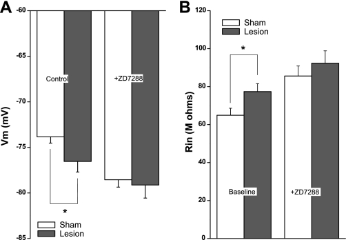
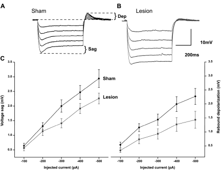
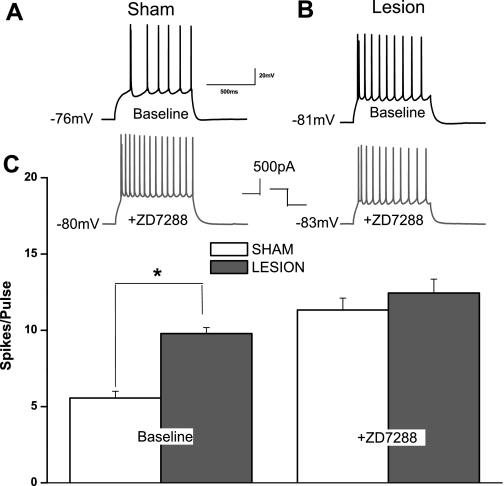
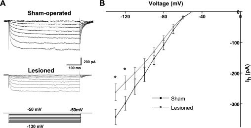
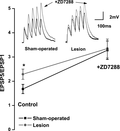
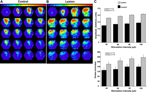
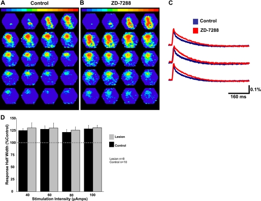
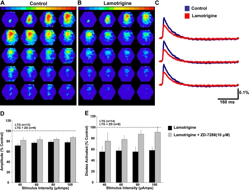
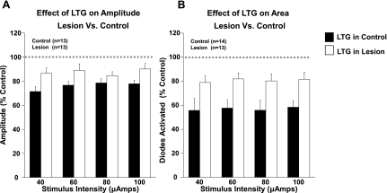
Focal cortical dysplasia is associated with the development of seizures in children and is present in up to 40% of intractable childhood epilepsies. Transcortical freeze lesions in newborn rats reproduce many of the anatomical and physiological characteristics of human cortical dysplasia. Rats with freeze lesions have increased seizure susceptibility and a region of hyperexcitable cortex adjacent to the lesion. Since alterations in hyperpolarization-activated nonspecific cation (HCN) channels are often associated with epilepsy, we used whole cell patch-clamp recording and voltage-sensitive dye imaging to examine alterations in HCN channels and inwardly rectifying hyperpolarization-activated currents (I(h)) in cortical dysplasia. (L5) pyramidal neurons in lesioned animals had hyperpolarized resting membrane potentials, increased input resistances and reduced voltage "sag" associated with I(h) activation. These differences became nonsignificant after application of the I(h) blocker ZD7288. Temporal excitatory postsynaptic potential (EPSP) summation and intrinsic excitability were increased in neurons near the freeze lesion. Using voltage-sensitive dye imaging of neocortical slices, we found that inhibiting I(h) with ZD7288 increased the half-width of dye signals. The anticonvulsant lamotrigine produced a significant decrease in spread of activity. The ability of lamotrigine to decrease network activity was reduced in the hyperexcitable cortex near the freeze lesion. These results suggest that I(h) serves to constrain network activity in addition to its role in regulating cellular excitability. Reduced I(h) may contribute to increased network excitability in cortical dysplasia.


Figures

Fig. 1.
Fig. 1.
L5 pyramidal neurons from freeze-lesioned rats have depolarized membrane potentials (Vm) and increased input resistances (Rin). A: the resting Vm of pyramidal neurons near the freeze lesion is significantly hyperpolarized compared with sham-operated control animals. This difference is not significant after hyperpolarization-activated nonspecific cation (HCN) channel inhibition with ZD7288. B: the Rin of pyramidal neurons near the freeze lesion is significantly higher than that of sham-operated control animals. This difference is not significant after HCN channel inhibition. *P < 0.05.
Fig. 2.
Fig. 2.
Reduction in hyperpolarization-activated current Ih-dependent voltage changes in L5 pyramidal neurons from lesioned animals. A: specimen records showing that membrane hyperpolarization in sham-operated animals is associated with a depolarizing “sag” in membrane voltage caused by Ih activation. Rebound depolarizations (Dep) are also seen. B: superimposed specimen records showing that sag responses are reduced in a pyramidal neuron near the freeze lesion. Rebound depolarizations upon current offset are also reduced. C: summary graphs showing a significant reduction in the amplitude in the voltage sag (left) and rebound depolarization (right) in pyramidal neurons from freeze-lesioned animals.
Fig. 3.
Fig. 3.
L5 pyramidal neurons from freeze-lesioned rats have increased intrinsic excitability. A: recordings showing a somatically evoked train of action potentials in a neuron from a sham-operated animal (top). In the same cell during bath application of ZD7288, Vm is hyperpolarized and the number of action potentials is increased (bottom). B: records obtained from a pyramidal neuron near a lesion. The same current injection resulted in a greater number of spikes under control conditions (top). After ZD7288, Vm and number of evoked action potentials are virtually unchanged. C: summary graphs showing difference in number of action potentials (APs) between sham-operated and lesioned animals before (left) and during (right) ZD7288. The difference in AP number is not significant after Ih inhibition. *P < 0.05.
Fig. 4.
Fig. 4.
Voltage-clamp recordings of Ih in neurons from sham-operated and lesioned animals. A, top: ZD7288-sensitive somatic Ih currents obtained by subtracting obtained before and after bath application of ZD7288 in a L5 pyramidal neurons from a sham-operated animal. Slowly activating Ih currents are observed. Bottom: recordings from a neuron near the freeze lesion revealed a significant decrease in Ih amplitude following membrane hyperpolarization. B: summary diagram showing current-voltage plots for a group of neurons in sham-operated and lesioned animals. *P < 0.05.
Fig. 5.
Fig. 5.
Effects of ZD7288 on excitatory postsynaptic potential (EPSP) summation in sham-operated and lesioned animals. Top left: specimen records of EPSPs evoked by a train of stimuli at 20 Hz. In a slice from a sham-operated animal, under control conditions EPSPs show weak facilitation. After ZD7288 amplitudes of EPSPs in this neuron were increased. Top right: similar experiment in a neuron from a slice from a lesioned animal. EPSPs evoked at 20 Hz summated to a significantly greater degree in pyramidal neurons from freeze-lesioned rats. EPSPs showed increased facilitation in presence of ZD7288. Bottom: graph of EPSP5-to-EPSP1 ratios shows that under control conditions ratios were significantly higher in sham-operated group. This difference was not significant after ZD7288. *P < 0.05.
Fig. 6.
Fig. 6.
Voltage-sensitive dye imaging of evoked activity. A: photograph showing the typical position of the brain slice over the diode array. The red dots indicate the borders of the hexagonal photodiode array. The array was positioned so that the upper limit was approximately in line with the pial surface. The arrow indicates the approximate position of the stimulating electrode. B: similar picture showing the typical position of the array relative to the freeze lesion. Small arrow indicates location of lesion. C: a pseudocolored image of peak activity is shown superimposed on the image of a slice. D: typical responses from selected individual diodes showing time course of fluorescence change.
Fig. 7.
Fig. 7.
Comparison of voltage-sensitive dye signals in control and lesioned animals. A: specimen record of a typical network response evoked from control cortex. B: a typical network response evoked in the hyperexcitable region adjacent to the freeze lesion. Evoked activity near the malformation in freeze-lesioned rats spreads further and is of higher amplitude. C: summary diagrams showing differences in response amplitude (top) and number of diodes activated (bottom) in lesioned vs. control animals.
Fig. 8.
Fig. 8.
HCN channel inhibition increases the duration of evoked network activity. A: typical network response evoked before HCN channel inhibition in a control animal. B: the same response after HCN channel inhibition with 10 μM ZD7288. C: responses from individual diodes before (blue) and after (red) HCN channel inhibition are shown superimposed. HCN channel inhibition increased the half-width of these responses. D: bar graphs showing that Ih inhibition increases the duration of evoked activity in both control and lesioned animals.
Fig. 9.
Fig. 9.
Effects of the anticonvulsant lamotrigine on evoked network activity. A: typical network response evoked in a control animal before lamotrigine. B: response to the same stimulation 20 min after application of lamotrigine. C: responses from individual diodes before (blue) and after (red) lamotrigine. D: lamotrigine (LTG) reduced the amplitude of diode responses in control in control animals. This effect was significantly attenuated by coapplication of ZD7288 (ZD). E: lamotrigine reduced the number of diodes activated (indicating spread of activity) in control animals. This effect was also significantly attenuated by coapplication of ZD7288.
Fig. 10.
Fig. 10.
Sensitivity of network activity to lamotrigine. A: the ability of lamotrigine (LTG) to decrease voltage-sensitive dye signal amplitude was significantly reduced in lesioned animals (open bars). B: the ability of lamotrigine to reduce the spread of the voltage-sensitive dye signal was significantly reduced in lesioned animals (open bars).

References

    1. Agmon A, Wells JE. The role of the hyperpolarization-activated cationic current Ih in the timing of interictal bursts in the neonatal hippocampus. J Neurosci 23: 3658–3668, 2003 - PMC - PubMed
    1. Aponte Y, Lien CC, Reisinger E, Jonas P. Hyperpolarization-activated cation channels in fast-spiking interneurons of rat hippocampus. J Physiol 574: 229–243, 2006 - PMC - PubMed
    1. Avanzini G, Franceschetti S, Mantegazza M. Epileptogenic channelopathies: experimental models of human pathologies. Epilepsia 48: 51–64, 2007 - PubMed
    1. Bandyopadhyay S, Hablitz JJ. NR2B antagonists restrict spatiotemporal spread of activity in a rat model of cortical dysplasia. Epilepsy Res 72: 127–139, 2006 - PubMed
    1. Becker AJ, Pitsch J, Sochivko D, Opitz T, Staniek M, Chen CC, Campbell KP, Schoch S, Yaari Y, Beck H. Transcriptional upregulation of Cav3.2 mediates epileptogenesis in the pilocarpine model of epilepsy. J Neurosci 28: 13341–13353, 2008 - PMC - PubMed

Publication types

MeSH terms

LinkOut - more resources