

Effectiveness of computer-aided detection in community mammography practice
- PMID: 21795668
- PMCID: PMC3149041
- DOI: 10.1093/jnci/djr206
Effectiveness of computer-aided detection in community mammography practice
Abstract
Background: Computer-aided detection (CAD) is applied during screening mammography for millions of US women annually, although it is uncertain whether CAD improves breast cancer detection when used by community radiologists.
Methods: We investigated the association between CAD use during film-screen screening mammography and specificity, sensitivity, positive predictive value, cancer detection rates, and prognostic characteristics of breast cancers (stage, size, and node involvement). Records from 684 956 women who received more than 1.6 million film-screen mammograms at Breast Cancer Surveillance Consortium facilities in seven states in the United States from 1998 to 2006 were analyzed. We used random-effects logistic regression to estimate associations between CAD and specificity (true-negative examinations among women without breast cancer), sensitivity (true-positive examinations among women with breast cancer diagnosed within 1 year of mammography), and positive predictive value (breast cancer diagnosed after positive mammograms) while adjusting for mammography registry, patient age, time since previous mammography, breast density, use of hormone replacement therapy, and year of examination (1998-2002 vs 2003-2006). All statistical tests were two-sided.
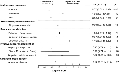
Results: Of 90 total facilities, 25 (27.8%) adopted CAD and used it for an average of 27.5 study months. In adjusted analyses, CAD use was associated with statistically significantly lower specificity (OR = 0.87, 95% confidence interval [CI] = 0.85 to 0.89, P < .001) and positive predictive value (OR = 0.89, 95% CI = 0.80 to 0.99, P = .03). A non-statistically significant increase in overall sensitivity with CAD (OR = 1.06, 95% CI = 0.84 to 1.33, P = .62) was attributed to increased sensitivity for ductal carcinoma in situ (OR = 1.55, 95% CI = 0.83 to 2.91; P = .17), although sensitivity for invasive cancer was similar with or without CAD (OR = 0.96, 95% CI = 0.75 to 1.24; P = .77). CAD was not associated with higher breast cancer detection rates or more favorable stage, size, or lymph node status of invasive breast cancer.
Conclusion: CAD use during film-screen screening mammography in the United States is associated with decreased specificity but not with improvement in the detection rate or prognostic characteristics of invasive breast cancer.
Figures
Comment in
-
Computer-assisted detection and screening mammography: where's the beef?J Natl Cancer Inst. 2011 Aug 3;103(15):1139-41. doi: 10.1093/jnci/djr267. Epub 2011 Jul 27. J Natl Cancer Inst. 2011. PMID: 21795666 No abstract available.
-
Re: effectiveness of computer-aided detection in community mammography practice.J Natl Cancer Inst. 2012 Jan 4;104(1):77; author reply 78-9. doi: 10.1093/jnci/djr492. Epub 2011 Dec 20. J Natl Cancer Inst. 2012. PMID: 22186178 Free PMC article. No abstract available.
-
Re: effectiveness of computer-aided detection in community mammography practice.J Natl Cancer Inst. 2012 Jan 4;104(1):77-8; author reply 78-9. doi: 10.1093/jnci/djr491. Epub 2011 Dec 20. J Natl Cancer Inst. 2012. PMID: 22186179 No abstract available.
References
-
- Chan HP, Doi K, Vyborny CJ, Lam KL, Schmidt RA. Computer-aided detection of microcalcifications in mammograms. Methodology and preliminary clinical study. Invest Radiol. 1988;23(9):664–671. - PubMed
-
- Rao VM, Levin DC, Parker L, Cavanaugh B, Frangos AJ, Sunshine JH. How widely is computer-aided detection used in screening and diagnostic mammography? J Am Coll Radiol. 2010;7(10):802–805. - PubMed
-
- Harvey D. Navigating through the data—CAD applications in liver, prostate, breast, and lung cancer. Radiol Today. 2010;11(3):18.
-
- Birdwell RL. The preponderance of evidence supports computer-aided detection for screening mammography. Radiology. 2009;253(1):9–16. - PubMed
-
- Philpotts LE. Can computer-aided detection be detrimental to mammographic interpretation? Radiology. 2009;253(1):17–22. - PubMed
Publication types
MeSH terms
Grants and funding
- U01 CA063740/CA/NCI NIH HHS/United States
- U01CA69976/CA/NCI NIH HHS/United States
- U01 CA070040/CA/NCI NIH HHS/United States
- U01CA70013/CA/NCI NIH HHS/United States
- U01 CA086082/CA/NCI NIH HHS/United States
- U01CA63736/CA/NCI NIH HHS/United States
- U01CA86082/CA/NCI NIH HHS/United States
- K05 CA104699/CA/NCI NIH HHS/United States
- CA104699/CA/NCI NIH HHS/United States
- U01CA70040/CA/NCI NIH HHS/United States
- U01CA63740/CA/NCI NIH HHS/United States
- U01 CA063731/CA/NCI NIH HHS/United States
- U01 CA086076/CA/NCI NIH HHS/United States
- U01 CA069976/CA/NCI NIH HHS/United States
- U01CA86076/CA/NCI NIH HHS/United States
- U01 CA063736/CA/NCI NIH HHS/United States
- U01 CA070013/CA/NCI NIH HHS/United States
- U01CA63731/CA/NCI NIH HHS/United States
LinkOut - more resources
Full Text Sources
Other Literature Sources
Medical
Miscellaneous
