

Comprehensive analysis of pathogen-specific antibody response in vivo based on an antigen library displayed on surface of yeast
- PMID: 21795672
- PMCID: PMC3190923
- DOI: 10.1074/jbc.M111.270553
Comprehensive analysis of pathogen-specific antibody response in vivo based on an antigen library displayed on surface of yeast
Abstract
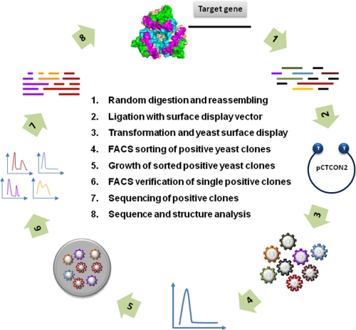
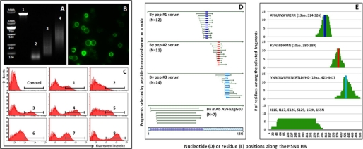
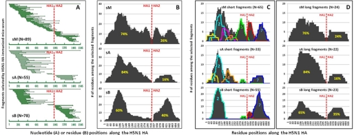
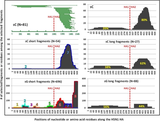
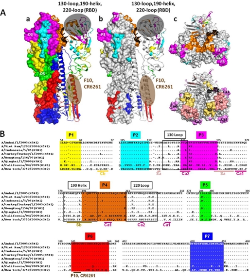
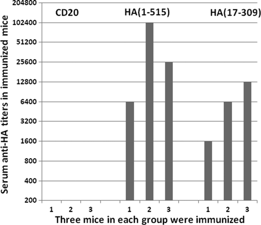
Host antibody response is a crucial defense against pathogenic infection. Here, we report a novel technique allowing quantitative measurement of polyclonal antibody response in vivo. This involves expression of a combinatorial library of target proteins from a candidate pathogen on the surface of yeast Saccharomyces cerevisiae. After mixing with serum/plasma from infected or immunized subjects, positive yeast clones were isolated via fluorescence-activated cell sorting (FACS). Using this technique, we have studied mouse immunized serum with recombinant hemagglutinin (HA) protein from a human influenza H5N1 strain (A/Anhui/1/2005) and convalescent plasma from an infected human in China. Our technique has identified novel antigenic domains targeted by serum/plasma and allowed calculation of the relative proportion of the antibody response against each domain. We believe such systematic measurement of an antibody response is unprecedented, and applying this method to different pathogens will improve understanding of protective immunity and guide development of vaccines and therapeutics.
Figures
Similar articles
-
Heterosubtypic neutralizing monoclonal antibodies cross-protective against H5N1 and H1N1 recovered from human IgM+ memory B cells.PLoS One. 2008;3(12):e3942. doi: 10.1371/journal.pone.0003942. Epub 2008 Dec 16. PLoS One. 2008. PMID: 19079604 Free PMC article.
-
Screening of random peptide library of hemagglutinin from pandemic 2009 A(H1N1) influenza virus reveals unexpected antigenically important regions.PLoS One. 2011 Mar 18;6(3):e18016. doi: 10.1371/journal.pone.0018016. PLoS One. 2011. PMID: 21437206 Free PMC article.
-
Potential Role of Nonneutralizing IgA Antibodies in Cross-Protective Immunity against Influenza A Viruses of Multiple Hemagglutinin Subtypes.J Virol. 2020 Jun 1;94(12):e00408-20. doi: 10.1128/JVI.00408-20. Print 2020 Jun 1. J Virol. 2020. PMID: 32269119 Free PMC article.
-
The antigenic architecture of the hemagglutinin of influenza H5N1 viruses.Mol Immunol. 2013 Dec;56(4):705-19. doi: 10.1016/j.molimm.2013.07.010. Epub 2013 Aug 7. Mol Immunol. 2013. PMID: 23933511 Review.
-
One step closer to universal influenza epitopes.Expert Rev Anti Infect Ther. 2009 Aug;7(6):687-90. doi: 10.1586/eri.09.48. Expert Rev Anti Infect Ther. 2009. PMID: 19681695 Review.
Cited by
-
Epitope-focused immunogens against the CD4-binding site of HIV-1 envelope protein induce neutralizing antibodies against auto- and heterologous viruses.J Biol Chem. 2018 Jan 19;293(3):830-846. doi: 10.1074/jbc.M117.816447. Epub 2017 Nov 29. J Biol Chem. 2018. PMID: 29187598 Free PMC article.
-
Unraveling of a neutralization mechanism by two human antibodies against conserved epitopes in the globular head of H5 hemagglutinin.J Virol. 2013 Mar;87(6):3571-7. doi: 10.1128/JVI.01292-12. Epub 2012 Dec 26. J Virol. 2013. PMID: 23269809 Free PMC article.
-
A human antibody recognizing a conserved epitope of H5 hemagglutinin broadly neutralizes highly pathogenic avian influenza H5N1 viruses.J Virol. 2012 Mar;86(6):2978-89. doi: 10.1128/JVI.06665-11. Epub 2012 Jan 11. J Virol. 2012. PMID: 22238297 Free PMC article.
-
Characterization of a monoclonal antibody against CREPT, a novel protein highly expressed in tumors.Monoclon Antib Immunodiagn Immunother. 2014 Dec;33(6):401-8. doi: 10.1089/mab.2014.0043. Monoclon Antib Immunodiagn Immunother. 2014. PMID: 25545209 Free PMC article.
-
Monoclonal antibodies specific to human Δ42PD1: A novel immunoregulator potentially involved in HIV-1 and tumor pathogenesis.MAbs. 2015;7(3):620-9. doi: 10.1080/19420862.2015.1016695. MAbs. 2015. PMID: 25692916 Free PMC article.
References
-
- Janeway C., Jr., Travers P., Walport M., Shlomchik M. (2001) in Immunobiology (Murphy M. K., Travers P. eds) Garland Science, New York
-
- Burton D. R. (2002) Nat. Rev. Immunol. 2, 706–713 - PubMed
-
- Dörner T., Radbruch A. (2007) Immunity 27, 384–392 - PubMed
-
- Scheid J. F., Mouquet H., Feldhahn N., Seaman M. S., Velinzon K., Pietzsch J., Ott R. G., Anthony R. M., Zebroski H., Hurley A., Phogat A., Chakrabarti B., Li Y., Connors M., Pereyra F., Walker B. D., Wardemann H., Ho D., Wyatt R. T., Mascola J. R., Ravetch J. V., Nussenzweig M. C. (2009) Nature 458, 636–640 - PubMed
Publication types
MeSH terms
Substances
Grants and funding
LinkOut - more resources
Full Text Sources
Other Literature Sources
Medical
