. 2011 Sep 23;286(38):33447-56.
doi: 10.1074/jbc.M111.225565. Epub 2011 Jul 27.

WW domain-containing E3 ubiquitin protein ligase 1 (WWP1) delays cellular senescence by promoting p27(Kip1) degradation in human diploid fibroblasts

Affiliations

WW domain-containing E3 ubiquitin protein ligase 1 (WWP1) delays cellular senescence by promoting p27(Kip1) degradation in human diploid fibroblasts

Xiaoxiao Cao et al. J Biol Chem. .

Abstract

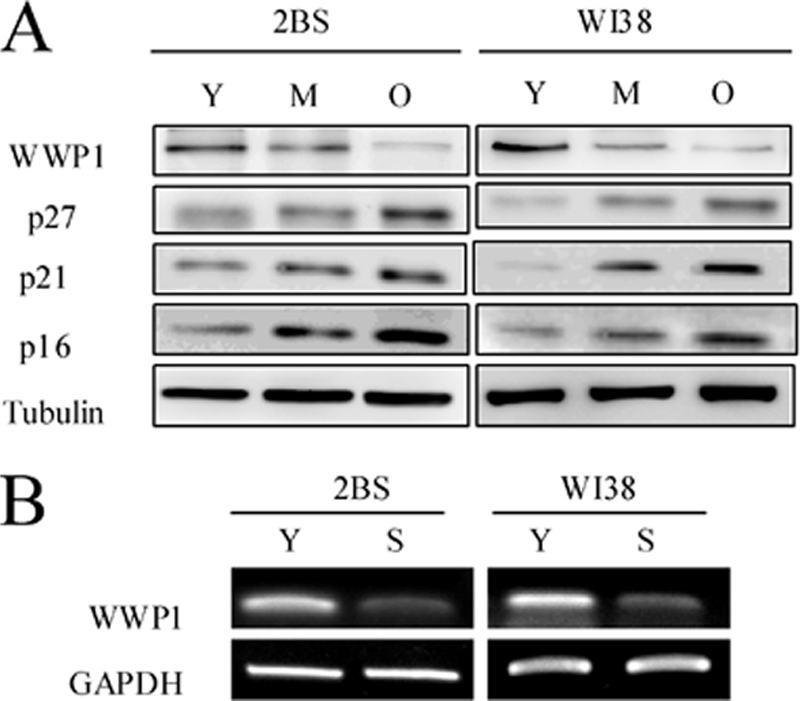
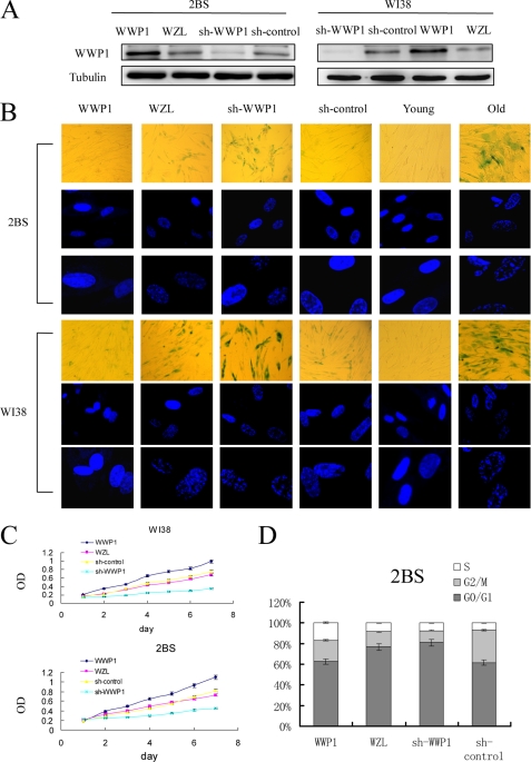
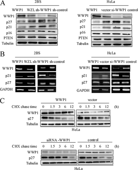
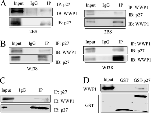
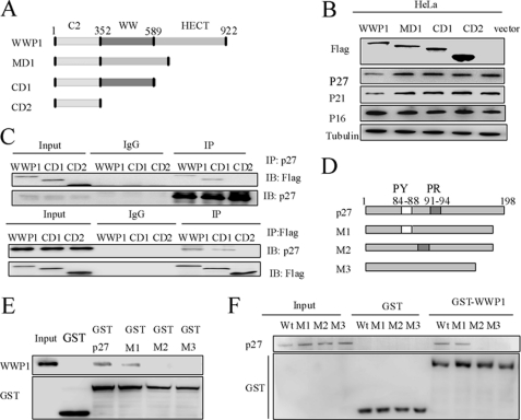
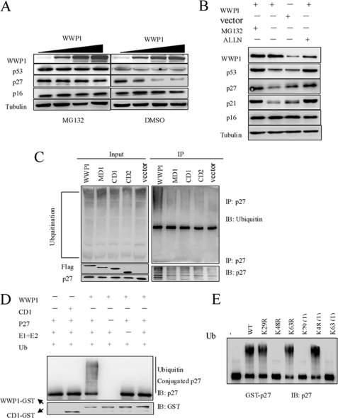
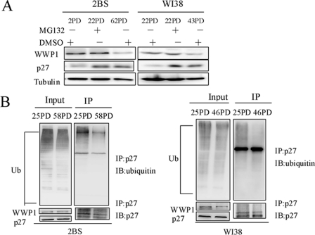
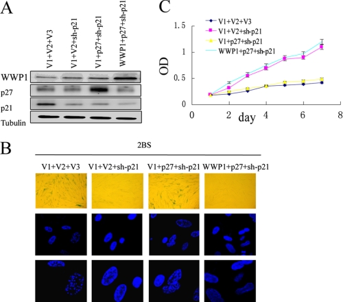
WW domain-containing E3 ubiquitin protein ligase 1 (WWP1) plays an important role in the proliferation of tumor cells and the lifespan of Caenorhabditis elegans. However, the role of WWP1 in cellular senescence is still unknown. Here, we show that the expression patterns of p27(Kip1) and WWP1 are inversely correlated during cellular senescence. Moreover, the overexpression of WWP1 delayed senescence, whereas the knockdown of WWP1 led to premature senescence in human fibroblasts. Furthermore, we demonstrate that WWP1 repressed endogenous p27(Kip1) expression through ubiquitin-proteasome-mediated degradation. Additionally, WWP1 had a strong preference for catalyzing the Lys-48-linked polyubiquitination of p27(Kip1) in vitro. Finally, we demonstrate that WWP1 markedly inhibited the replicative senescence induced by p27(Kip1) by promoting p27(Kip1) degradation. Therefore, our study provides a new molecular mechanism for the regulation of cellular senescence.


Figures

FIGURE 1.
FIGURE 1.
Expression patterns of WWP1 in young, middle-aged, and senescent cells. A, Western blot analysis of WWP1, p16 INK4a, p21Cip1, and p27Kip1 expression in young (Y), middle-aged (M), and senescent (O) 2BS and WI38 cells. Total protein was extracted, and immunoblotting was performed using specific antibodies against WWP1, p16INK4a, p21Cip1, and p27Kip1 as indicated. Tubulin served as a loading control. B, RT-PCR analysis of WWP1 in young (Y) and senescent (S) 2BS and WI38 cells. Total mRNA was extracted and assessed by RT-PCR using specific primers. GAPDH was used as a loading control.
FIGURE 2.
FIGURE 2.
WWP1 repressed senescence-associated features in 2BS and WI38 cells. The stable transformants were passaged until senescence was achieved. They were then analyzed for the related senescence markers. A, Western blot analysis of WWP1 expression levels in WWP1-infected or WWP1 shRNA-infected cells. B, WWP1-transfected and WWP1 shRNA-transfected 2BS and WI38 cells were stained for SA-β-gal activity (upper panel) and formation of SAHF (middle and lower panels). C, growth curves of WWP1-transfected and WWP1 shRNA-transfected cells were determined by the MTT assay. Values are the mean ± S.D. of triplicate points from a representative experiment (n = 3), which was repeated three times with similar results. D, flow cytometry analysis of WWP1-transfected and WWP1 shRNA-transfected 2BS cells. Values represent the means ± S.E. of triplicate points from a representative experiment (n = 3), which was repeated three times. sh, shRNA.
FIGURE 3.
FIGURE 3.
WWP1 decreased p27Kip1 protein levels. A, Western blot analysis of WWP1, p21Cip1, p27Kip1, and p16INK4a expression was carried out in WWP1-overexpressing or knockdown cells compared with control cells. 2BS cells and HeLa cells were used for these experiments. B, RT-PCR analysis of WWP1, p21Cip1, and p27Kip1 was performed in cells as in A. Specific RT-PCR primers for WWP1, p21Cip1, and p27Kip1 were used. C, the half-life of p27Kip1 was evaluated in HeLa cells with altered WWP1 expression. Forty-eight hours post-transfection, cells were treated with cycloheximide (CHX) (15 μg/ml) for different time periods. Blots were evaluated with WWP1, p27Kip1, and tubulin antibodies. sh, shRNA; si, siRNA.
FIGURE 4.
FIGURE 4.
WWP1 interacted with p27Kip1in vitro and in vivo. A and B, co-immunoprecipitation (IP) of endogenous WWP1 and p27Kip1 was performed in 2BS and WI38 cells. C, immunoprecipitation of WWP1 and p27Kip1 in WWP1-transfected HeLa cells using the p27Kip1 antibody. D, GST pulldown assay using in vitro transcribed and translated WWP1 and purified GST-p27. Blots were evaluated with WWP1 and GST antibodies. IB, immunoblot.
FIGURE 5.
FIGURE 5.
WWP1 associated with p27Kip1 directly through WW domain of WWP1 and LPEFY motif of p27Kip1. A, schematic representation of the WWP1 protein sequence and the fragments used in further assays. B, HeLa cells were transfected with FLAG-tagged WWP1, CD1, CD2, and MD1 and a vector control. After 48 h, cell lysates were processed for Western blot analysis of FLAG, p16INK4a, p21Cip1, and p27Kip1. C, cells were transfected with WWP1, CD1, and CD2 and treated with MG132 (15 μm) for 5 h before collection. Cell lysates were then used for co-immunoprecipitation (IP) with the p27Kip1 antibody or FLAG antibody. Blots were evaluated with FLAG and p27Kip1 antibodies. D, schematic representation of wild-type p27Kip1 and its deletions. E and F, GST pulldown assay of WWP1 and p27Kip1 or three deletions of p27Kip1, including M1, M2, and M3. Blots were evaluated with WWP1, p27Kip1, and GST antibodies. IB, immunoblot.
FIGURE 6.
FIGURE 6.
WWP1 polyubiquitinated p27Kip1. A, different amounts of WWP1 were expressed in HeLa cells. Twenty-four hours after transfection, cells were treated with MG132 (15 μm) for 5 h before collection. Proteins were analyzed by Western blotting. B, HeLa cells were transfected with WWP1 and vector. Twenty-four hours after transfection, WWP1-transfected cells were treated with MG132 (15 μm) or acetyl-Leu-Leu-norleucinal (ALLN) (20 μm) 5 h before harvesting. Proteins were analyzed by Western blotting. C, an in vivo ubiquitination assay was performed with WWP1 and p27Kip1. HeLa cells were transiently transfected with WWP1, CD1, CD2, and MD1 constructs. Forty-two hours later, cells were treated with MG132 (15 μm) for 5 h. Cells were lysed and processed for co-immunoprecipitation (IP) using the p27Kip1 antibody. Blots were analyzed with ubiquitin, p27Kip1, and FLAG antibodies. D, an in vitro ubiquitination assay was performed. Affinity-purified p27Kip1 was incubated with purified WWP1, E1 enzyme, UbcH7, and ubiquitin. The reaction mixture was incubated at 37 °C for 90 min and analyzed by Western blotting. E, in vitro ubiquitination of p27Kip1 was performed in the presence of human E1, UbcH7, and WWP1 with wild-type ubiquitin or the indicated ubiquitin (Ub) mutant proteins. Following a 90-min reaction, the products were analyzed by Western blotting. IB, immunoblot.
FIGURE 7.
FIGURE 7.
WWP1 expression positively correlated with p27Kip1 ubiquitination level during cellular senescence. A, Western blot analysis of the expression patterns of WWP1 and p27Kip1 in young cells (treated with MG132 or DMSO) and old cells (treated with DMSO). Tubulin served as a loading control. B, young human fibroblasts (22PD 2BS and 25PD WI38) and old human fibroblasts (58PD 2BS and 46PD WI38) were treated with MG132 (15 μm) for 5 h. Cells were lysed and processed for co-immunoprecipitation (IP) using the p27Kip1 antibody. Blots were analyzed with ubiquitin, p27Kip1, and WWP1 antibodies. IB, immunoblot.
FIGURE 8.
FIGURE 8.
WWP1 repressed cellular senescence induced by p27Kip1 in the presence of p21Cip1 inhibitor. A, the expression of WWP1, p27Kip1, and p21Cip1 protein levels in the stable transformants was analyzed by Western blotting. B, cells from A were stained for SA-β-gal activity (upper panel) and formation of SAHF (middle and lower panels). C, growth curves for cells from A were determined using the MTT assay. Values represent the mean ± S.D. of triplicate points from a representative experiment (n = 3), which was repeated three times with similar results. sh, shRNA.

References

    1. Bayreuther K., Rodemann H. P., Hommel R., Dittmann K., Albiez M., Francz P. I. (1988) Proc. Natl. Acad. Sci. U.S.A. 85, 5112–5116 - PMC - PubMed
    1. Dimri G. P., Lee X., Basile G., Acosta M., Scott G., Roskelley C., Medrano E. E., Linskens M., Rubelj I., Pereira-Smith O., Et A. (1995) Proc. Natl. Acad. Sci. U.S.A. 92, 9363–9367 - PMC - PubMed
    1. Narita M., Nũnez S., Heard E., Narita M., Lin A. W., Hearn S. A., Spector D. L., Hannon G. J., Lowe S. W. (2003) Cell 113, 703–716 - PubMed
    1. Schulz L., Tyler J. (2005) Mol. Cell 17, 168–170 - PubMed
    1. Bringold F., Serrano M. (2000) Exp. Gerontol. 35, 317–329 - PubMed

Publication types

MeSH terms

LinkOut - more resources