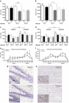
Decreased proliferation of adult hippocampal stem cells during cocaine withdrawal: possible role of the cell fate regulator FADD
- PMID: 21796105
- PMCID: PMC3176567
- DOI: 10.1038/npp.2011.119
Decreased proliferation of adult hippocampal stem cells during cocaine withdrawal: possible role of the cell fate regulator FADD
Abstract
The current study uses an extended access rat model of cocaine self-administration (5-h session per day, 14 days), which elicits several features manifested during the transition to human addiction, to study the neural adaptations associated with cocaine withdrawal. Given that the hippocampus is thought to have an important role in maintaining addictive behavior and appears to be especially relevant to mechanisms associated with withdrawal, this study attempted to understand how extended access to cocaine impacts the hippocampus at the cellular and molecular levels, and how these alterations change over the course of withdrawal (1, 14, and 28 days). Therefore, at the cellular level, we examined the effects of cocaine withdrawal on cell proliferation (Ki-67+ and NeuroD+ cells) in the DG. At the molecular level, we employed a 'discovery' approach with gene expression profiling in the DG to uncover novel molecules possibly implicated in the neural adaptations that take place during cocaine withdrawal. Our results suggest that decreased hippocampal cell proliferation might participate in the adaptations associated with drug removal and identifies 14 days as a critical time-point of cocaine withdrawal. At the 14-day time-point, gene expression profiling of the DG revealed the dysregulation of several genes associated with cell fate regulation, highlighting two new neurobiological correlates (Ascl-1 and Dnmt3b) that accompany cessation of drug exposure. Moreover, the results point to Fas-Associated protein with Death Domain (FADD), a molecular marker previously associated with the propensity to substance abuse and cocaine sensitization, as a key cell fate regulator during cocaine withdrawal. Identifying molecules that may have a role in the restructuring of the hippocampus following substance abuse provides a better understanding of the adaptations associated with cocaine withdrawal and identifies novel targets for therapeutic intervention.
Figures


Similar articles
-
Cocaine withdrawal causes delayed dysregulation of stress genes in the hippocampus.PLoS One. 2012;7(7):e42092. doi: 10.1371/journal.pone.0042092. Epub 2012 Jul 30. PLoS One. 2012. PMID: 22860061 Free PMC article.
-
Methamphetamine binge administration dose-dependently enhanced negative affect and voluntary drug consumption in rats following prolonged withdrawal: role of hippocampal FADD.Addict Biol. 2019 Mar;24(2):239-250. doi: 10.1111/adb.12593. Epub 2017 Dec 28. Addict Biol. 2019. PMID: 29282816
-
Hippocampal cell fate regulation by chronic cocaine during periods of adolescent vulnerability: Consequences of cocaine exposure during adolescence on behavioral despair in adulthood.Neuroscience. 2015 Sep 24;304:302-15. doi: 10.1016/j.neuroscience.2015.07.040. Epub 2015 Jul 26. Neuroscience. 2015. PMID: 26215918
-
Synaptic Microtubule-Associated Protein EB3 and SRC Phosphorylation Mediate Structural and Behavioral Adaptations During Withdrawal From Cocaine Self-Administration.J Neurosci. 2019 Jul 17;39(29):5634-5646. doi: 10.1523/JNEUROSCI.0024-19.2019. Epub 2019 May 15. J Neurosci. 2019. PMID: 31092585 Free PMC article.
-
The impact of cocaine on adult hippocampal neurogenesis: Potential neurobiological mechanisms and contributions to maladaptive cognition in cocaine addiction disorder.Biochem Pharmacol. 2017 Oct 1;141:100-117. doi: 10.1016/j.bcp.2017.05.003. Epub 2017 May 6. Biochem Pharmacol. 2017. PMID: 28483462 Review.
Cited by
-
Dose-Dependent Antidepressant-Like Effects of Cannabidiol in Aged Rats.Front Pharmacol. 2022 Jul 1;13:891842. doi: 10.3389/fphar.2022.891842. eCollection 2022. Front Pharmacol. 2022. PMID: 35847003 Free PMC article.
-
Cocaine-induced Changes in the Expression of NMDA Receptor Subunits.Curr Neuropharmacol. 2019;17(11):1039-1055. doi: 10.2174/1570159X17666190617101726. Curr Neuropharmacol. 2019. PMID: 31204625 Free PMC article. Review.
-
Effects of addictive drugs on adult neural stem/progenitor cells.Cell Mol Life Sci. 2016 Jan;73(2):327-48. doi: 10.1007/s00018-015-2067-z. Epub 2015 Oct 14. Cell Mol Life Sci. 2016. PMID: 26468052 Free PMC article. Review.
-
The Interplay between the Hippocampus and Amygdala in Regulating Aberrant Hippocampal Neurogenesis during Protracted Abstinence from Alcohol Dependence.Front Psychiatry. 2013 Jun 27;4:61. doi: 10.3389/fpsyt.2013.00061. Print 2013. Front Psychiatry. 2013. PMID: 23818882 Free PMC article.
-
Self administration of oxycodone alters synaptic plasticity gene expression in the hippocampus differentially in male adolescent and adult mice.Neuroscience. 2015 Jan 29;285:34-46. doi: 10.1016/j.neuroscience.2014.11.013. Epub 2014 Nov 14. Neuroscience. 2015. PMID: 25446355 Free PMC article.
References
-
- Ahmed SH, Koob GF. Transition from moderate to excessive drug intake: change in hedonic set point. Science. 1998;282:298–300. - PubMed
-
- Altman J, Das GD. Autoradiographic and histological evidence of postnatal hippocampal neurogenesis in rats. J Comp Neurol. 1965;124:319–335. - PubMed
-
- Alvaro-Bartolome M, Esteban S, García-Gutiérrez MS, Manzanares J, Valverde O, García-Sevilla JA. Regulation of Fas receptor/Fas-associated protein with death domain apoptotic complex and associated signalling systems by cannabinoid receptors in the mouse brain. Br J Pharmacol. 2010;160:643–656. - PMC - PubMed
-
- Arguello AA, Harburg GC, Schonborn JR, Mandyam CD, Yamaguchi M, Eisch AJ. Time course of morphine's effects on adult hippocampal subgranular zone reveals preferential inhibition of cell in S phase of the cell cycle and a subpopulation of immature neurons. Neuroscience. 2008;157:70–79. - PMC - PubMed
-
- Banasr M, Soumier A, Hery M, Mocaër E, Daszuta A. Agomelatine, a new antidepressant, induces regional changes in hippocampal neruogenesis. Biol Psychiatry. 2006;59:1087–1096. - PubMed
Publication types
MeSH terms
Substances
Grants and funding
LinkOut - more resources
Full Text Sources
Medical
Research Materials
Miscellaneous

