Review
. 2011 Sep;20 Suppl 5(Suppl 5):686-98.
doi: 10.1007/s00586-011-1937-9. Epub 2011 Jul 28.

Hip-spine relations and sagittal balance clinical consequences

Affiliations
Review

Hip-spine relations and sagittal balance clinical consequences

Jean-Yves Lazennec et al. Eur Spine J. 2011 Sep.

Abstract

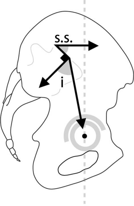
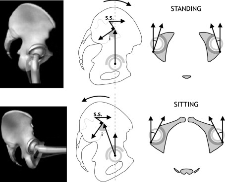
Introduction: The role of the pelvic area in sagittal balance is evident for spinal surgeons, but the influence of the coxofemoral joint is underestimated and inadequately explained by conventional imagery. Comprehensive analysis of the pelvic and subpelvic sectors as part of the sagittal, frontal and cross-sectional balance of the trunk sheds new light on some spinal diseases and their relation to the pelvis.

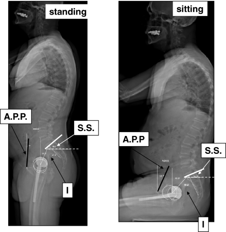
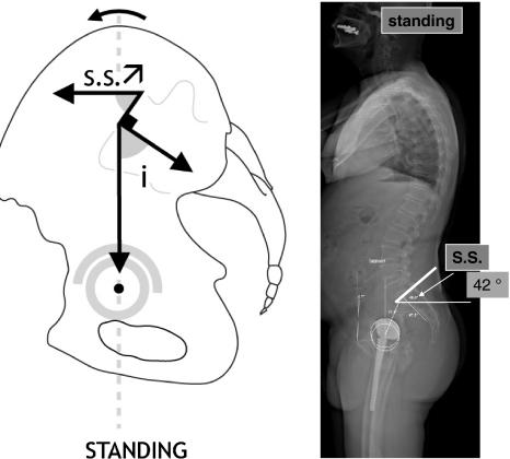
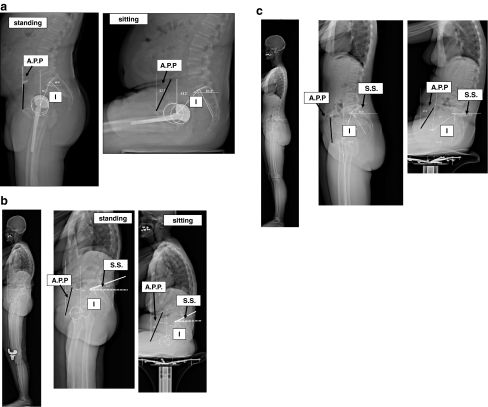
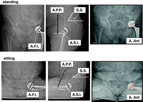
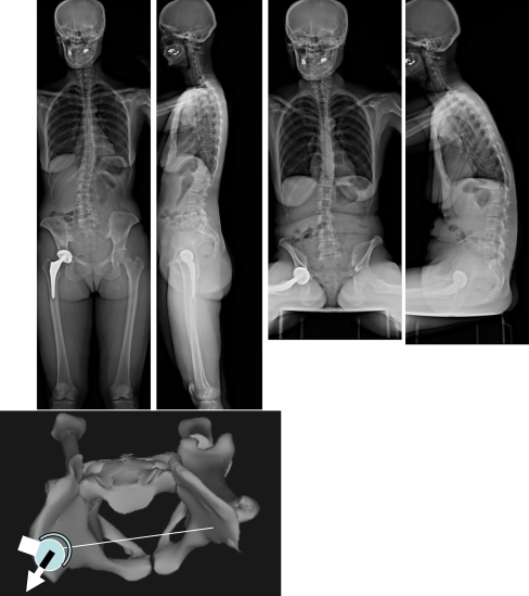
Methods: This analysis, based on innovative radiologic methods as the EOS(®) technology but also on a new look at conventional imaging makes it possible to better analyze standing lateral images and seated images.

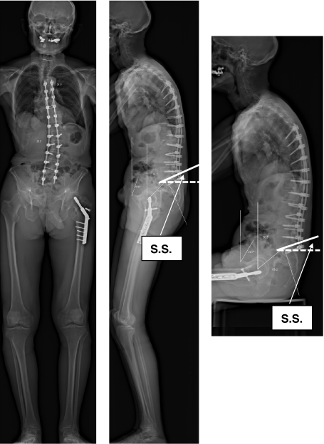
Results: Disturbances can come from atypical morphotypes or from unusual postures as in aging spine. The measurement of available extension and the concept of available flexion provide new information regarding individual's adaptation to the imbalance induced by disorders of the spine or lower limbs.

Conclusion: A comprehensive assessment of each patient and in particular of the complex comprising the spine and the pelvis, is essential for understanding each individual's adaptation to the imbalance induced by disorders of the spine or lower limbs.


Figures

Fig. 1
Fig. 1
Definition of the pelvic incidence (morphologic parameter invariant for a given subject, with a mean value of 50–55°) and of the sacral slope (functional parameter that varies according to position)
Fig. 2
Fig. 2
EOS® views of the sagittal balance of a patient with a THA, in standing and seated positions. The incidence does not change regardless of position. The sacral slope is higher in a standing than a seated position. Simultaneous variations of the inclination of Lewinnek’s plane (the posterior tilt of this plane is associated with the posterior pelvic tilt in a seated position). I incidence, SS sacral slope, APP angle between the Lewinnek plane and the vertical plane
Fig. 3
Fig. 3
Diagram of the standing position: anterior tilt (flexion) of the pelvis with horizontalization of the sacrum and increase of the sacral slope. The incidence is invariable and does not change
Fig. 4
Fig. 4
Permanent posterior pelvic tilt and imbalance of a spine stiffened by a long arthrodesis. The sacral slope is low in both the standing and seated positions. In standing position, the pelvis and the spine function as if the patient were in a seated position
Fig. 5
Fig. 5
Patient with an exaggerated anterior pelvic tilt: the sacrum is very horizontal in standing position with a sacral slope greater than normal
Fig. 6
Fig. 6
Diagram of the seated position: posterior pelvic tilt with verticalization of the sacrum and decrease of the sacral slope. The incidence is invariable and does not change
Fig. 7
Fig. 7
Variations in sagittal balance from the standing to the seated position. a Usual variation: The anterior pelvic plane tilts backwards in a seated position at the same time that the sacral slope is reduced from 45° standing to 26° seated; in this case, the available pelvic flexion is 19°. b Little postural variation of a stiffened lumbosacral joint (no available pelvic flexion). c Strong postural variations, sacral slope negative in a seated position (available pelvic flexion 38°)
Fig. 8
Fig. 8
Modification of the acetabular orientation from standing to seated position: increase in the acetabular frontal inclination (AFI.), the acetabular sagittal inclination (ASI), and the acetabular anterior opening (A Ant) measured by radiologic anteversion (Pradhan)
Fig. 9
Fig. 9
The unusual posterior tilt of the pelvis can be detected on the AP standing image by the overly visible foramina obturata. After THA, this could result in a posterior impingement in standing position (anterior dislocation or subluxation)
Fig. 10
Fig. 10
Principle of measurement of the available spinopelvic extension (EOS® system). a The patient is assessed in a lateral standing position. (b, c) Each hip is then assessed in its maximum range of extension in standing position, placing the contralateral lower limb on a step such that it is in a position of maximum flexion of the coxofemoral joint. The extrinsic available extension is measured by the capacity to increase the sacral slope (SS 2–SS 1). The intrinsic available extension is measured by the variation of the sacro-femoral angle (SFA). The sacro-femoral angle is defined as the angle between the segment drawn from the middle of the sacral plate to the middle of the segment joining the centers of the femoral heads, and the segment joining the latter point (midpoint of the segment joining the center of the femoral heads) to the summit of the Blumensaat line, at the knee. The intrinsic extension is 177 − 152° = 25° for the THA side and 177 − 158° = 19° for the non-operated hip
Fig. 11
Fig. 11
a Situation of spinopelvic imbalance and hips that do not extend completely. The incidence is high; it is theoretically associated with elevated lordosis to keep the sagittal balance stable. In this case, the patient has an anterior imbalance not compensated by the hips (absence of hyperextension). This imbalance can be analyzed by searching for the available extension. b No role for the spine in recovering the extension: no available extrinsic extension because there is no modification of the sacral slope. There is no available extension for the non-operated hip (full extension truly impossible). Only the hip that underwent THA has an increase in available extension: from 164 − 146° = 18°. The presence of this available extension on the operated side is evidence of an adaptive false inextendability in the reference standing position
Fig. 12
Fig. 12
The abnormal anterior tilt of the pelvis in a seated position can lead to an anterior impingement of the hips with the risk of lesions of the labrum and the cartilage. In these cases, the available hip flexion required by the seated position is exaggerated, as the pelvic posterior tilt (pelvic retroversion) is not sufficient
Fig. 13
Fig. 13
Incidence is a morphologic characteristic determinant in the adaptation of the sagittal spinopelvic balance. In subjects with a high angle of pelvic incidence, lumbar lordosis is greater and the range of adaptation of the sacral slope may be greater. The anterior opening of the acetabulum is less marked, and the available extension of the hips greater. In the case of low pelvic incidence, lumbar lordosis is lower and the range of adaptation of the sacral slope more limited. The anterior opening of the acetabulum is more marked, but the theoretically available extension is lower at the hips
Fig. 14
Fig. 14
In standing position, the sacral slope is high (40° approximately); the acetabulum appears more horizontal from both frontal and lateral views; the functional acetabular anteversion is weaker. In a seated position, the sacral slope is lower, sometimes negative, and the acetabulum appears more vertical from both the frontal and lateral views. The functional acetabular anteversion is more elevated
Fig. 15
Fig. 15
Modifications of the functional mobility cone in cases of pelvic tilt disorders: in the case of a posterior tilt of the pelvis, the cone is deflected; even though the coxofemoral joints are perfectly mobile, the patient is using his available extension
Fig. 16
Fig. 16
Substantial rotation of the pelvis associated with scoliosis: the THA is perfectly implanted relative to the bone landmarks but subluxated permanently forward in the standing position: pelvic rotation induced an abnormal acetabular anteversion in a standing position. Note that the pelvic rotation disappears in the seated position (patient is asymptomatic when sitting)

References

    1. Kobayashi T, et al. A longitudinal study of congruent sagittal spinal alignment in an adult cohort. Spine (Phila Pa 1976) 2004;29(6):671–676. doi: 10.1097/01.BRS.0000115127.51758.A2. - DOI - PubMed
    1. Chanplakorn P, et al. Lumbopelvic alignment on standing lateral radiograph of adult volunteers and the classification in the sagittal alignment of lumbar spine. Eur Spine J. 2011;20(5):706–712. doi: 10.1007/s00586-010-1626-0. - DOI - PMC - PubMed
    1. Vialle R, et al. Radiographic analysis of the sagittal alignment and balance of the spine in asymptomatic subjects. J Bone Joint Surg Am. 2005;87(2):260–267. doi: 10.2106/JBJS.D.02043. - DOI - PubMed
    1. Jackson RP, McManus AC. Radiographic analysis of sagittal plane alignment and balance in standing volunteers and patients with low back pain matched for age, sex, and size. A prospective controlled clinical study. Spine (Phila Pa 1976) 1994;19(14):1611–1618. doi: 10.1097/00007632-199407001-00010. - DOI - PubMed
    1. Padgett DE, Warashina H (2004) The unstable total hip replacement. Clin Orthop Relat Res 420(1):72–79 - PubMed