Review
. 2011 Sep;20 Suppl 5(Suppl 5):626-33.
doi: 10.1007/s00586-011-1930-3. Epub 2011 Jul 28.

Sagittal balance disorders in severe degenerative spine. Can we identify the compensatory mechanisms?

Affiliations
Review

Sagittal balance disorders in severe degenerative spine. Can we identify the compensatory mechanisms?

Cédric Barrey et al. Eur Spine J. 2011 Sep.

Abstract

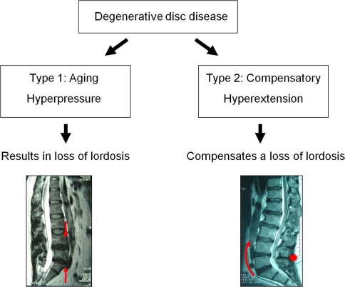
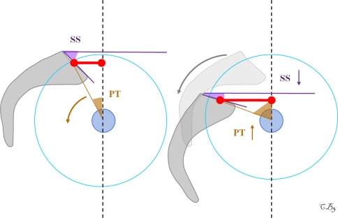
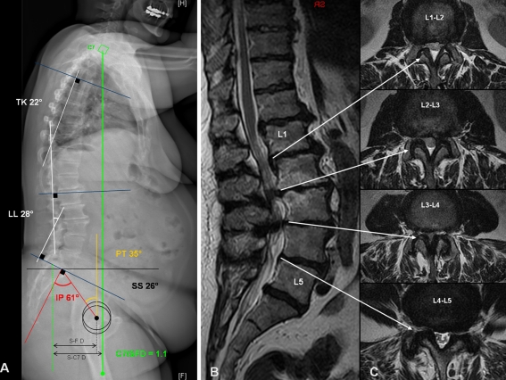
Introduction: Aging of the spine is characterized by facet joints arthritis, degenerative disc disease and atrophy of extensor muscles resulting in a progressive kyphosis. Recent studies confirmed that patients with lumbar degenerative disease were characterized by an anterior sagittal imbalance, a loss of lumbar lordosis and an increase of pelvis tilt. The aim of this paper was thus to describe the different compensatory mechanisms which are observed in the spine, pelvis and/or lower limbs areas for patients with severe degenerative spine.

Methods: We reviewed all the compensatory mechanisms of sagittal unbalance described in the literature.

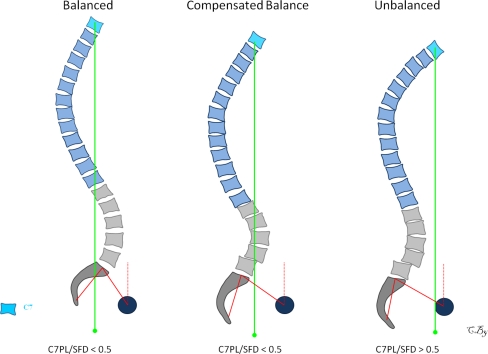
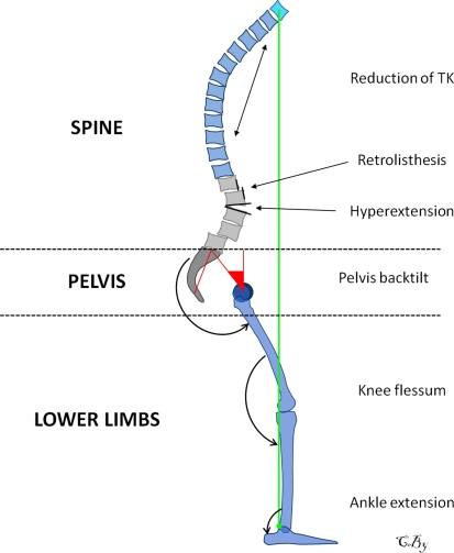
Results: According to the severity of the imbalance, we could identify three different stages: balanced, balanced with compensatory mechanisms and imbalanced. For the two last stages, the compensatory mechanisms permitted to limit consequences of lumbar kyphosis on the global sagittal alignment. Reduction of thoracic kyphosis, intervertebral hyperextension, retrolisthesis, pelvis backtilt, knee flessum and ankle extension were the main mechanisms described in the literature. The basic concept of these compensatory mechanisms was to extend adjacent segments of the kyphotic spine allowing for compensation of anterior translation of the axis of gravity.

Conclusions: To avoid underestimate the severity of the degenerative spine disorder, it thus seems important to recognize the different compensatory mechanisms from the upper part of the trunk to the lower limbs. We propose a three steps algorithm to analyse the balance status and determine the presence or not of these compensatory mechanisms: measurement of pelvis incidence, assessment of global sagittal alignment and analysis of compensatory mechanisms successively in the spine, pelvis and lower limbs areas.


Figures

Fig. 1
Fig. 1
Evaluation of global sagittal alignment using the spino-sacral angle (a) and the C7/SFD ratio (b). The SSA is defined as the angle between the sacral plate and the line connecting the centroid of C7 vertebral body and the midpoint of the sacral plate [23]. Sacro-femoral distance (SFD) is the horizontal distance between the vertical bi-coxo-femoral axis and the vertical line passing through the posterior corner of the sacrum. The horizontal distance between C7 PL and the posterior corner of the sacrum (that is SC7 D) was also measured. Then we calculated the C7/SFD ratio corresponding to the ratio between SC7 Distance and SF Distance [1]
Fig. 2
Fig. 2
Classification of global sagittal alignment in 3 stages with respect to the severity of the imbalance: stage 1 balanced, stage 2 balanced with compensatory mechanisms, stage 3 unbalanced (C7PL/SFD ratio superior to 0.5)
Fig. 3
Fig. 3
Sagittal imbalance and the different compensatory mechanisms in the spine, pelvis and lower limbs areas
Fig. 4
Fig. 4
Patient with lumbar kyphosis and severe multilevel stenosis from L2–L3 to L4–L5: full spine radiographs (a), sagittal T2-weighted (b) and transverse T2-weighted (c) MRI sequences. The patient is still balanced (C7PL/SFD is 0.25), but balance is compensated by three main mechanisms: pelvis back tilt (curved arrow), multilevel retrolisthesis (red circles) and reduction of thoracic kyphosis (calculated to 25°). PI was measured to 47°, PT was 34° and SS was 13°. Compared to group control from normal and asymptomatic population, we should expect value of PT around 10°. On MRI axial slices, retrolisthesis at L3–L4 and L4–L5 are associated with fluid collection in facet joints (straight arrows)
Fig. 5
Fig. 5
Patient with lumbar stenosis from L2–L3 to L4–L5 and thoraco-lumbar kyphosis: full spine radiographs (a), X-rays focused on lumbo-pelvic zone (b) and sagittal T2-weighted MRI sequence (c). The patient is well-balanced (C7PL/SFD is −0.3) however, some compensatory mechanisms are present in the lumbar area. Hyperextension is observed at L5–S1 (curvedblack arrow) (local lordosis was measured to 24°) and there are multilevel retrolisthesis at L2–L3 (red circles) and L4–L5 (large arrow). The pelvis tilt was quite normal as it was calculated to 22° and the PI to 46°
Fig. 6
Fig. 6
Classification of degenerative disc diseases into aging discopathy and compensatory discopathy
Fig. 7
Fig. 7
Pelvis back tilt mechanism. Increase of pelvis tilt results in posterior placement of sacrum related to the coxo-femoral heads thus increasing the sacro-femoral distance (red lines)
Fig. 8
Fig. 8
Patient with multilevel lumbar stenosis from L1–L2 to L4–L5: full spine radiographs (a), sagittal T2-weighted MRI sequence (b) and axial T2-weighted MRI sequences from L1–L2 to L4–L5 (c). The patient is clearly unbalanced (SC7D/SFD is more than 1) with C7 plumbline in front of femoral heads. The sagittal unbalance is because of the lumbar kyphosis, which is secondary to the severe degenerative disc diseases from T12 to S1. Seeing that PI is measured to 61° and compared to group control, we should expect value of LL around 70° (only measured to 28° on full spine radiographs). To limit sagittal unbalance, this patient undergoes pelvis retroversion (increase of PT which is calculated to more than 30°), reduction of thoracic kyphosis (only 22°) and knee flessum

References

    1. Barrey C (2004) Equilibre sagittal pelvi-rachidien et pathologies lombaires dégénératives. Etude comparative à propos de 100 cas (in French). Thèse de Médecine. Université Claude Bernard, Lyon
    1. Barrey C, Jund J, Noseda O, Roussouly P. Sagittal balance of the pelvis–spine complex and lumbar degenerative diseases. A comparative study about 85 cases. Eur Spine J. 2007;16:1459–1467. doi: 10.1007/s00586-006-0294-6. - DOI - PMC - PubMed
    1. Barrey C, Jund J, Perrin G, Roussouly P. Spinopelvic alignment of patients with degenerative spondylolisthesis. Neurosurg. 2007;61:981–986. doi: 10.1227/01.neu.0000303194.02921.30. - DOI - PubMed
    1. Berthonnaud E, Dimnet J, Roussouly P, Labelle H. Analysis of the sagittal spine and pelvis using shape and orientation parameters. J Spinal Disord Tech. 2005;18:40–47. doi: 10.1097/01.bsd.0000117542.88865.77. - DOI - PubMed
    1. During J, Goudfrooij H, Keessen W, Beeker TW, Crowe A. Towards standards for posture. Postural characteristics of the lower back system in normal and pathologic conditions. Spine. 1985;10:83–87. doi: 10.1097/00007632-198501000-00013. - DOI - PubMed

MeSH terms