. 2012 Jul 1;131(1):30-40.
doi: 10.1002/ijc.26323. Epub 2011 Aug 25.

Inhibition of sonic hedgehog pathway and pluripotency maintaining factors regulate human pancreatic cancer stem cell characteristics

Affiliations

Inhibition of sonic hedgehog pathway and pluripotency maintaining factors regulate human pancreatic cancer stem cell characteristics

Su-Ni Tang et al. Int J Cancer. .

Abstract

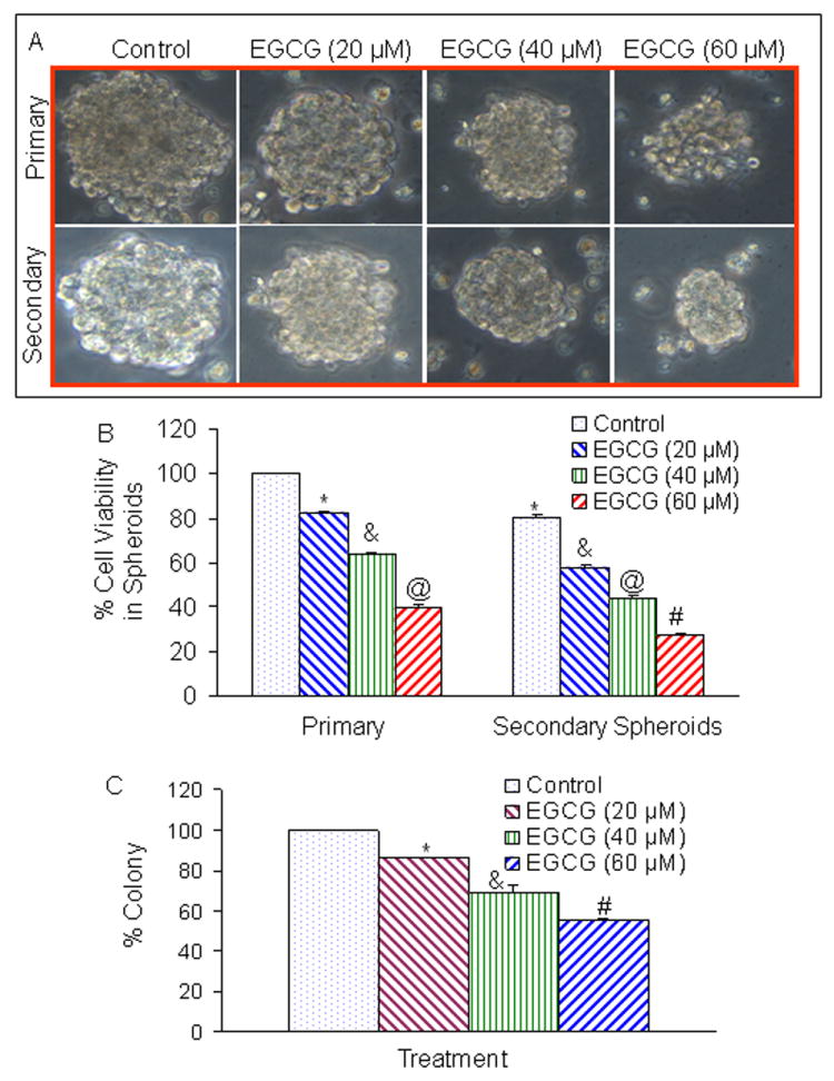
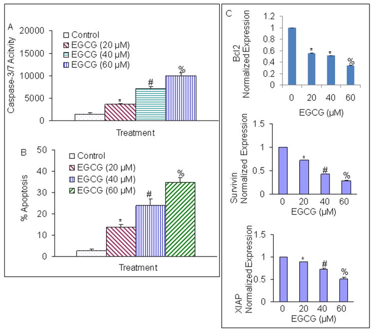
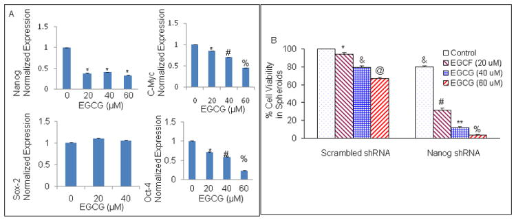
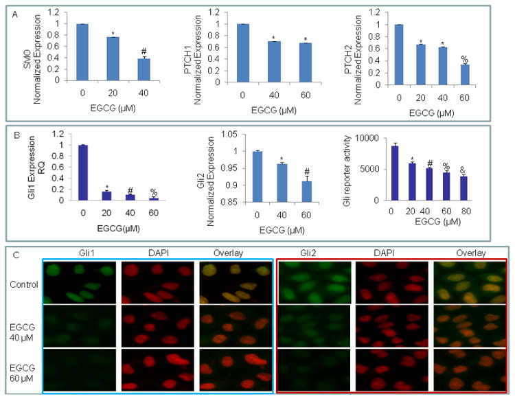
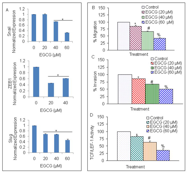
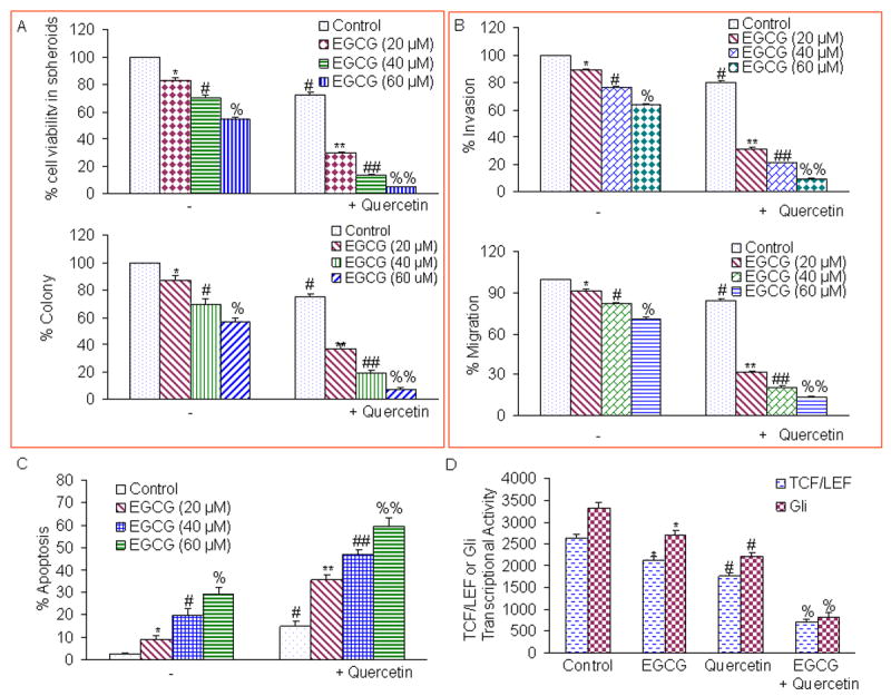
Activation of the sonic hedgehog (SHh) pathway is required for the growth of numerous tissues and organs and recent evidence indicates that this pathway is often recruited to stimulate growth of cancer stem cells (CSCs) and to orchestrate the reprogramming of cancer cells via epithelial mesenchymal transition (EMT). The objectives of this study were to examine the molecular mechanisms by which (-)-epigallocatechin-3-gallate (EGCG), an active compound in green tea, inhibits self-renewal capacity of pancreatic CSCs and synergizes with quercetin, a major polyphenol and flavonoid commonly detected in many fruits and vegetables. Our data demonstrated that EGCG inhibited the expression of pluripotency maintaining transcription factors (Nanog, c-Myc and Oct-4) and self-renewal capacity of pancreatic CSCs. Inhibition of Nanog by shRNA enhanced the inhibitory effects of EGCG on self-renewal capacity of CSCs. EGCG inhibited cell proliferation and induced apoptosis by inhibiting the expression of Bcl-2 and XIAP and activating caspase-3. Interestingly, EGCG also inhibited the components of SHh pathway (smoothened, patched, Gli1 and Gli2) and Gli transcriptional activity. Furthermore, EGCG inhibited EMT by inhibiting the expression of Snail, Slug and ZEB1, and TCF/LEF transcriptional activity, which correlated with significantly reduced CSC's migration and invasion, suggesting the blockade of signaling involved in early metastasis. Furthermore, combination of quercetin with EGCG had synergistic inhibitory effects on self-renewal capacity of CSCs through attenuation of TCF/LEF and Gli activities. Since aberrant SHh signaling occurs in pancreatic tumorigenesis, therapeutics that target SHh pathway may improve the outcomes of patients with pancreatic cancer by targeting CSCs.


Conflict of interest statement

Disclosure of Potential Conflicts of Interest

The authors indicate no potential conflicts of interest.

Figures

Fig. 1
Fig. 1
Effects of EGCG on tumor spheroids and cell viability of pancreatic cancer stem cells (CSCs). (A), Pancreatic CSCs were seeded in suspension and treated with EGCG (0-60 μM) for 7 days. Pictures of spheroids formed in suspension were taken by a microscope. (B), Pancreatic CSCs were seeded in suspension and treated with EGCG (0-60 μM) for 7 days. At the end of incubation period, sheroids were collected, and dissociated with Accutase (Innovative Cell Technologies, Inc.). For secondary spheroids, cells were reseeded and treated with EGCG (0-60 μM) for 7 days. Cell viability was measured by trypan blue assay. Data represent mean ± SD. *, &, @, or # = significantly different from respective controls, P < 0.05. (C), EGCG inhibits colony formation by CSCs. Pancreatic CSCs were seeded in soft agar and treated with various doses of EGCG and incubated at 4°C for 21 days. At the end of incubation period, colonies were counted. Data represent mean ± SD. *, & or # = significantly different from respective controls, P < 0.05.
Fig. 2
Fig. 2
Regulation of caspase-3/7 activity, apoptosis and apoptosis-related proteins by EGCG on CSCs derived from human primary pancreatic tumors. (A), Regulation of caspase-3/7 activity by EGCG. CSCs were treated with EGCG (0-60 μM) for 24 h, and caspase-3/7 activity was measured as per manufacturer’s instructions. Data represent mean ± SD. *, # or % = significantly different from control, P < 0.05. (B), Regulation of apoptosis by EGCG. CSCs were treated with EGCG (0-60 μM) for 48 h, and apoptosis was measured by TUNEL assay. Data represent mean ± SD. *, # or % = significantly different from control, P < 0.05. (C), Regulation of apoptosis-related proteins. Pancreatic CSCs were treated with EGCG (0-60 μM) for 36 h. Real time PCR (q-RT-PCR) was performed to examine the expression of Bcl-2, survivin, XIAP, and GAPDH. Data represent mean ± SD. *, # or % = significantly different from control, P < 0.05.
Fig. 3
Fig. 3
Regulation of pluripotency maintaining transcription factors by EGCG in pancreatic cancer stem cells. (A) Pancreatic CSCs were treated with EGCG (0-60 μM) for 36 h. At the end of incubation period, cells were harvested and the expression of Nanog, Sox-2, c-Myc and Oct-4 was measured by the q-RT-PCR. Data represent mean ± SD. *, #, or % = significantly different from respective controls, P < 0.05. (B), Nanog shRNA enhances the inhibitory effects of EGCG on CSC’s spheroid viability. Pancreatic CSCs were transduced with either scrambled shRNA or Nanog shRNA expressing lentiviral vector (pLKO.1), and cell lysates were collected and western blot analysis was performed using anti-Nanog antibody (data not shown). CSC/scrambled and CSC/Nanog shRNA were seeded as described above and treated with EGCG (0-60 μM). After 7 days, spheroids were collected and cell suspensions were prepared and viable cells were counted by trypan blue assay. Data represent mean ± SD. *, &, @, #, ** or % = significantly different from control, P < 0.05.
Fig. 4
Fig. 4
Inhibition of components of sonic hedgehog pathway, Gli transcription and nuclear translocation by EGCG. (A) Inhibition of components of sonic hedgehog pathway and Gli transcription. Pancreatic CSCs were treated with EGCG (0-60 μM) for 36 h. The expression of Smothened (Smo), patched 1 (PTCH1), patched 2 (PTCH2), was measured by q-RT-PCR. Data represent mean ± SD. *, #, or % = significantly different from respective controls, P < 0.05. (B), Inhibition of Gli1 and Gli2 expression and Gli transcription. Pancreatic CSCs were treated with EGCG (0-60 μM) for 36 h. The expression of Gli1 and Gli2 was measured by q-RT-PCR. Gli reporter activity. CSCs were transduced with Gli-responsive GFP/firefly luciferase viral particles (pGreen Fire1-Gli with EF1, System Biosciences). After transduction, culture medium was replaced and CSCs were treated with EGCG (0-60 μM) for 24 h. Gli-responsive reporter activity was measured by luciferase assay (Promega Corporation). Data represent mean ± SD. *, #, % or & = significantly different from respective controls, P < 0.05. (C), EGCG inhibits nuclear translocation of Gli1 and Gli2. Pancreatic CSCs were treated with or without EGCG (40 or 60 μM) for 24 h. At the end of incubation period, CSCs were fixed with paraformaldehyde, permeabilized with titron X100, and blocked with 5% normal goat serum. Cells were then treated with either anti-Gli1 or anti-Gli2 antibody, followed by secondary antibody plus DAPI. Stained cells were mounted and visualized under a fluorescence microscope. Blue fluorescence of DAPI was changed to red color for a better contrast.
Fig. 5
Fig. 5
Regulation of epithelial mesenchymal transition factors, migration, invasion and TCF/LEF activity by EGCG in pancreatic CSCs. (A), Pancreatic CSCs were treated with EGCG (0-60 μM) for 48 h. At the end of incubation period, the expression of Snail, ZEB1 and Slug was measured by q-RT-PCR. Data represent mean ± SD. * = significantly different from respective controls, P < 0.05. (B), Transwell migration assay. Pancreatic CSCs were plated in the top chamber of the transwell and treated with EGCG (0-60 μM) for 24 h. Cells migrated to the lower chambered were fixed with methanol, stained with crystal violet and counted. Data represent mean ± SD. * # or % = significantly different from respective controls, P < 0.05. (C) Matrigel invasion assay. CSCs were plated onto the Matrigel-coated membrane in the top chamber of the transwell and treated with EGCG (0-60 μM) for 48 h. Cells invaded to the lower chambered were fixed with methanol, stained with crystal violet and counted. Data represent mean ± SD. *, # or % = significantly different from respective controls, P < 0.05. (D), Effects of EGCG on TCF-1/LEF activity. Pancreatic CSCs were transduced with TCF/LEF responsive GFP/firefly luciferase viral particles (pGreen Fire1-Gli with EF1, System Biosciences). Transduced CSCs were treated with EGCG (0-60) for 48 h and the GFP fluorescence was measured. Data represent mean ± SD. *, # or ** = significantly different from control, P < 0.05.
Fig. 6
Fig. 6
Quercetin synergizes with EGCG to inhibit self-renewal capacity, invasion, migration, and TCF/LEF and Gli transcriptional activities in pancreatic CSCs. (A), Effects of EGCG and quercetin on spheroid and colony formation. Upper Panel, Quercetin synergizes with EGCG to inhibit spheroid’s cell viability. CSCs were seeded in suspension and treated with EGCG (0-60 μM) with or without quercetin (20 μM) for 7 days. At the end of incubation period, all the spheroids were collected and resuspended. Cell viability was measured by trypan blue assay. Data represent mean ± SD. *, #, %, **, ##, or %% = significantly different from control, P < 0.05. Lower panel, Quercetin synergizes with EGCG to inhibit colony formation. Pancreatic CSCs were seeded in soft agar and treated with various doses of EGCG (0-60 μM) with or without quercetin (20 μM) and incubated at 4°C for 21 days. At the end of incubation period, colonies were counted. Data represent mean ± SD. *, #, %, **, ##, or %% = significantly different from control, P < 0.05. (B), Effects of EGCG and quercetin on invasion and migration. Upper panel, Matrigel invasion assay. CSCs were plated onto the Matrigel-coated membrane in the top chamber of the transwell and treated with EGCG (0-60 μM) with or without quercetin (20 μM) for 48 hrs. Cells invaded to the lower chambered were fixed with methanol, stained with crystal violet and counted. Data represent mean ± SD. *, #, %, **, ##, or %% = significantly different from control, P < 0.05. Lower panel, Transwell migration assay. Pancreatic CSCs were plated in the top chamber of the transwell and treated with EGCG (0-60 μM) with or without quercetin (20 μM) for 48 hrs. Cells migrated to the lower chambered were fixed with methanol, stained with crystal violet and counted. Data represent mean ± SD. *, #, %, **, ##, or %% = significantly different from respective controls, P < 0.05. (C), Quercetin synergizes with EGCG to induce apoptosis. CSCs were seeded in suspension and treated with EGCG (0-60 μM) with or without quercetin (20 μM) for 7 days. At the end of incubation period, all the spheroids were collected. Apoptosis was measured by TUNEL assay. Data represent mean ± SD. *, #, %, **, ##, or %% = significantly different from control, P < 0.05. (D), Effects of EGCG and quercetin on TCF/LEF and Gli transcriptional activities. Pancreatic CSCs were transduced with either lentivirus encoding TCF/LEF responsive GFP and luciferase genes or Gli-responsive GFP and luciferase genes. Transduced CSCs were treated with EGCG (40 μM) with or without quercetin (20 μM) for 48 hrs and the luciferase activity was measured. Data represent mean ± SD. *, #, or % = significantly different from respective control, P < 0.05.

Similar articles

Cited by

References

    1. Jemal A, Siegel R, Xu J, Ward E. Cancer statistics, 2010. CA Cancer J Clin. 2010;60:277–300. - PubMed
    1. Pliarchopoulou K, Pectasides D. Pancreatic cancer: current and future treatment strategies. Cancer Treat Rev. 2009;35:431–6. - PubMed
    1. Magee CJ, Ghaneh P, Neoptolemos JP. Surgical and medical therapy for pancreatic carcinoma. Best Pract Res Clin Gastroenterol. 2002;16:435–55. - PubMed
    1. Yeo TP, Hruban RH, Leach SD, Wilentz RE, Sohn TA, Kern SE, Iacobuzio-Donahue CA, Maitra A, Goggins M, Canto MI, Abrams RA, Laheru D, et al. Pancreatic cancer. Curr Probl Cancer. 2002;26:176–275. - PubMed
    1. Warshaw AL, Fernandez-del Castillo C. Pancreatic carcinoma. N Engl J Med. 1992;326:455–65. - PubMed

Publication types

MeSH terms