

doi: 10.1002/cbic.201100046.
Epub 2011 Jul 27.
Identifying protein variants with cross-reactive aptamer arrays
Affiliations
- PMID: 21796750
- PMCID: PMC3454492
- DOI: 10.1002/cbic.201100046
Item in Clipboard
Identifying protein variants with cross-reactive aptamer arrays
Chembiochem.
.
No abstract available
Figures
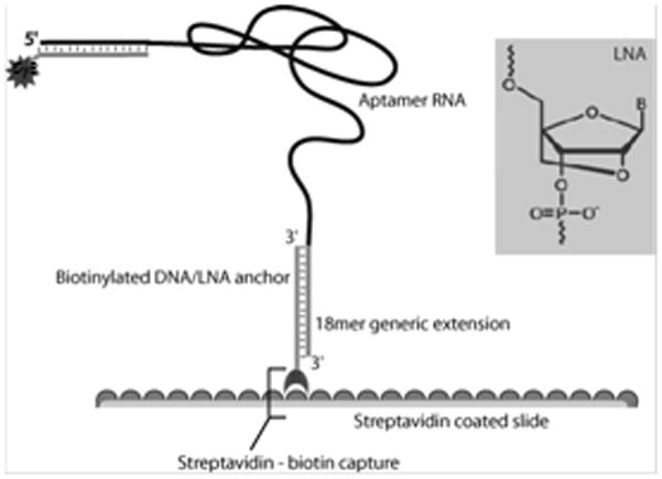
A biotinylated LNA anchor complementary to the 5′ end of the aptamer, securing the aptamer to the Nutravidin coated slide. A second LNA conjugated to the 3′ end of the aptamer acts as a probe for detection of bound aptamer.
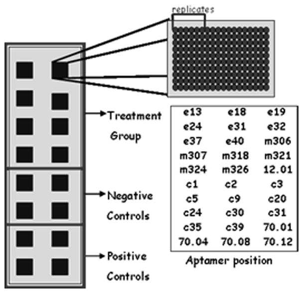
Slide treatment. Each small square represents a single reaction well, 16 per slide. Each well was identical and consisted of 30 aptamers printed in replicates of 6. The aptamer’s position is a representation of where each aptamer was positioned relative to the others; each name represents a set of six replicates. Each slide was separated into three groups of wells. The top eight wells correspond to the treatment group; where one of the four HIV-RT variants were applied. The next four wells correspond to negative controls and the final four correspond to the positive controls.
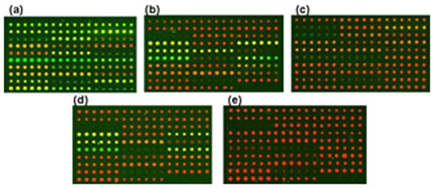
GenePix scan of the 30 aptamer set. Red corresponds to the Cy5 channel and the signal intensity is proportional to the amount of aptamer bound to the slide. “Black” spots indicated locations where little or no aptamer was deposited. If the background intensity exceeded the foreground intensity in either channel the spots were excluded. Green corresponds to the Cy3 channel where the signal intensity is proportional to the amount of protein bound to the aptamer. a) Wild-type b) M3 c) M5 d) M9 e) negative control.
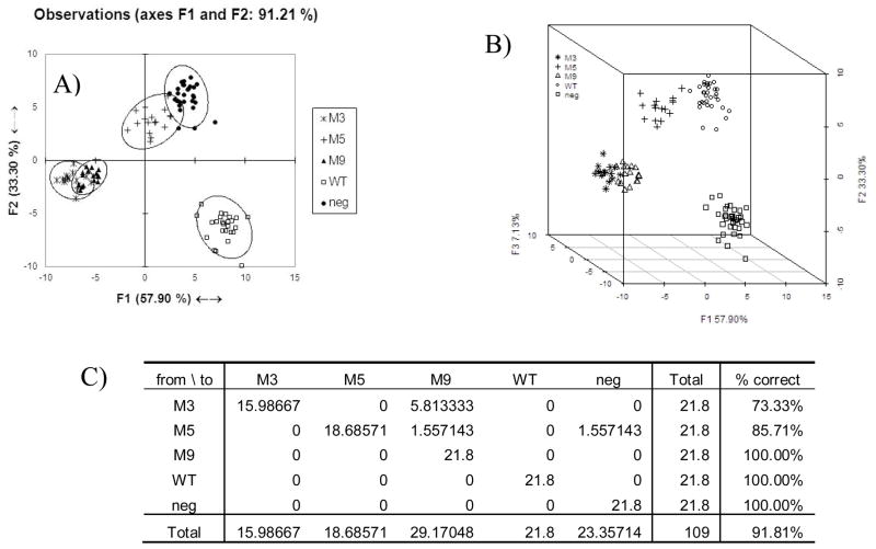
a)“ within-slide” normalized LDA of 30 aptamers set 91.2% explained. Ellipses represent 95% confidence intervals b) LDA of 30 aptamer set including 3 components “within slide normalized”, 98.33% captured. c) Leave-one out cross validation
Similar articles
-
Isolation of Peptide aptamers to target protein function.Methods Mol Biol. 2009;535:333-60. doi: 10.1007/978-1-59745-557-2_19. Methods Mol Biol. 2009. PMID: 19377987
-
Prediction of aptamer-protein interacting pairs using an ensemble classifier in combination with various protein sequence attributes.BMC Bioinformatics. 2016 May 31;17(1):225. doi: 10.1186/s12859-016-1087-5. BMC Bioinformatics. 2016. PMID: 27245069 Free PMC article.
-
Development of an Aptamer-Based Lateral Flow Assay for the Detection of C-Reactive Protein Using Microarray Technology as a Prescreening Platform.ACS Comb Sci. 2020 Nov 9;22(11):617-629. doi: 10.1021/acscombsci.0c00080. Epub 2020 Sep 7. ACS Comb Sci. 2020. PMID: 32894679
-
Peptide aptamers as guides for small-molecule drug discovery.Drug Discov Today. 2006 Apr;11(7-8):334-41. doi: 10.1016/j.drudis.2006.02.007. Drug Discov Today. 2006. PMID: 16580975 Review.
-
Peptide aptamers: development and applications.Curr Top Med Chem. 2015;15(12):1082-101. doi: 10.2174/1568026615666150413153143. Curr Top Med Chem. 2015. PMID: 25866267 Free PMC article. Review.
Cited by
-
A differential fluorescent receptor for nucleic acid analysis.Chembiochem. 2014 Jan 24;15(2):228-31. doi: 10.1002/cbic.201300657. Epub 2013 Dec 11. Chembiochem. 2014. PMID: 24339354 Free PMC article.
-
Array-based "Chemical Nose" Sensing in Diagnostics and Drug Discovery.Angew Chem Int Ed Engl. 2019 Apr 8;58(16):5190-5200. doi: 10.1002/anie.201809607. Epub 2019 Feb 20. Angew Chem Int Ed Engl. 2019. PMID: 30347522 Free PMC article. Review.
-
Next-generation sequencing as input for chemometrics in differential sensing routines.Angew Chem Int Ed Engl. 2015 May 18;54(21):6339-42. doi: 10.1002/anie.201501822. Epub 2015 Mar 31. Angew Chem Int Ed Engl. 2015. PMID: 25826754 Free PMC article.
-
Fingerprinting Non-Terran Biosignatures.Astrobiology. 2018 Jul;18(7):915-922. doi: 10.1089/ast.2017.1712. Epub 2018 Mar 8. Astrobiology. 2018. PMID: 29634318 Free PMC article.
-
Small molecule detection with aptamer based lateral flow assays: Applying aptamer-C-reactive protein cross-recognition for ampicillin detection.Sci Rep. 2018 Apr 4;8(1):5628. doi: 10.1038/s41598-018-23963-6. Sci Rep. 2018. PMID: 29618771 Free PMC article.
References
-
- Conroy PJ, Hearty S, Leonard P, O’Kennedy RJ. Semin Cell Dev Biol. 2009;20:10–26. - PubMed
- Luppa PB, Sokoll LJ, Chan DW. Clin Chim Acta. 2001;314:1–26. - PubMed
- Goodey A, Lavigne JJ, Savoy SM, Rodriguez MD, Curey T, Tsao A, Simmons G, Wright J, Yoo SJ, Sohn Y, Anslyn EV, Shear JB, Neikirk DP, McDevitt JT. J AM CHEM SOC. 2001;123:2559–2570. - PubMed
- Haab BB, Dunham MJ, Brown PO. Genome Biol. 2001;2 RESEARCH0004. - PMC - PubMed
-
- Miller JC, Zhou H, Kwekel J, Cavallo R, Burke J, Butler EB, Teh BS, Haab BB. Proteomics. 2003;3:56–63. - PubMed
-
- Lavigne J, Anslyn E. ANGEW CHEM INT EDIT. 2001;40:3119–3130. - PubMed
- Dickinson TA, White J, Kauer JS, Walt DR. Nature. 1996;382:697–700. - PubMed
- Albert KJ, Lewis NS, Schauer CL, Sotzing GA, Stitzel SE, Vaid TP, Walt DR. CHEM REV. 2000;100:2595–2626. - PubMed
- Lewis NS. Acc Chem Res. 2004;37:663–672. - PubMed
-
- Shangguan D, Cao Z, Meng L, Mallikaratchy P, Sefah K, Wang H, Li Y, Tan W. J PROTEOME RES. 2008;7:2133–2139. - PMC - PubMed
- Suslick K, Rakow N, Sen A. TETRAHEDRON. 2004;60:11133–11138.
- Stojanovic M, Green E, Semova S, Nikic D, Landry D. J AM CHEM SOC. 2003;125:6085–6089. - PubMed
- Fitter S, James R. J BIOL CHEM. 2005;280:34193–34201. - PubMed
Publication types
MeSH terms
Substances
Grants and funding
LinkOut - more resources
Full Text Sources
Other Literature Sources
