. 2011 Nov;79(11):894-6.
doi: 10.1002/cyto.a.21112. Epub 2011 Jul 27.

OMIP-003: phenotypic analysis of human memory B cells

Affiliations

OMIP-003: phenotypic analysis of human memory B cells

Chungwen Wei et al. Cytometry A. 2011 Nov.
No abstract available


Figures

Figure 1
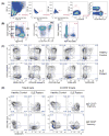
Figure 1. Classification of core human B cell subsets and phenotypic characterization of memory B cells in healthy subjects and patients with Systemic Lupus Erythematosus
(A) Lymphocytes gated through the FSC-A vs SSC-A plot were further interrogated by the ratios of Height to Width in forward scatter and side scatter, as well as their ability to uptake the amine-reactive Aqua fluorescent dye in order to gate out cell aggregates and dead cells, respectively. Live CD19+CD3 B cells were then selected for analysis as shown in B. (B) The customarily used IgD/CD27 scheme classifies peripheral blood B cells into four core subsets (left panel): naïve (N: IgD+CD27); unswitched memory (Um: IgD+CD27+); IgDCD27+ switched memory (CD27+ Sm); IgDCD27 switched memory B cells (CD27 Sm). IgDCD27++ plasmablasts (PB) are a rare population in steady-state healthy subjects and can be better discriminated as CD27++CD38++ cells in the IgD fraction of a healthy subject who, in this example, received HPV vaccination 11 days prior to the analysis (middle panel). The rightmost panel shows that autoreactive 9G4+ B cells concentrate within the naïve compartment in healthy subjects (3). The comparisons among different B cell classification schemes made possible by this panel is discussed and illustrated in Online Figure 3. (C) Comparisons of IgD B cells between a healthy subject and a SLE patient illustrate the value of multicolor panels to reveal informative phenotypic differences in health and disease. As previously reported (2), B220 expression is lost during GC differentiation and thus CD27+ resting memory cells are predominantly B220−. In contrast, IgDCD27 cells, which as in this example may be the dominant memory population in active SLE, are predominantly B220+. This expanded subset is also characterized in SLE by the down-regulation of CD24 and CD21, markers expressed by the majority of PBL B cells in general and CD27+ memory B cells in particular. Loss of CD21 and up-regulation of CD95 have been independently associated with memory B cell activation (4,5). Accordingly, expansions of activated switched memory subsets (CD27+ and CD27) are evident in active SLE. In contrast, CXCR3 expression (suggestive of migratory potential of activated cells to Th1 areas of systemic inflamed tissues) is concentrated in the IgDCD27+ memory subset. (D) The inclusion of CD21, CD95 and CXCR3 in the same panel allows informative co-localization of these markers. In contrast with CD27+ resting memory cells in the healthy control, a significant fraction of IgDCD27 cells lack expression of CD21, a feature consistent with activation. Yet, the lack of CD95 expression indicates that these two markers are not necessarily correlated. In SLE, CD95+ cells are greatly expanded in both memory subsets and the vast majority of IgDCD27CD95+ cells are also CD21. In contrast, CD95+ cells within the lupus IgDCD27+ memory are almost equally split between CD21+ and CD21, illustrating again that the expression of these markers is not necessarily reciprocal. The CXCR3+ fractions of both IgDCD27+ and IgDCD27 memory subsets exhibit CD21/CD95 expression patterns that reflect the differences generally observed between healthy subjects and SLE patients. As illustrated in this example, the expression of CD95 in IgDCD27CXCR3+ cells is increased in SLE.

References

    1. Wei C, Anolik J, Cappione A, Zheng B, Pugh-Bernard A, Brooks J, Lee E-H, Milner ECB, Sanz I. A New Population of Cells Lacking Expression of CD27 Represents a Notable Component of the B Cell Memory Compartment in Systemic Lupus Erythematosus. J Immunol. 2007;178(10):6624–6633. - PubMed
    1. Sanz I, Wei C, Lee FE-H, Anolik J. Phenotypic and functional heterogeneity of human memory B cells. Seminars in Immunology. 2008;20(1):67–82. - PMC - PubMed
    1. Cappione A, 3rd, Anolik JH, Pugh-Bernard A, Barnard J, Dutcher P, Silverman G, Sanz I. Germinal center exclusion of autoreactive B cells is defective in human systemic lupus erythematosus. J Clin Invest. 2005;115(11):3205–16. - PMC - PubMed
    1. Moir S, Ho J, Malaspina A, Wang W, DiPoto AC, O’Shea MA, Roby G, Kottilil S, Arthos J, Proschan MA, et al. Evidence for HIV-associated B cell exhaustion in a dysfunctional memory B cell compartment in HIV-infected viremic individuals. J Exp Med. 2008;205(8):1797–1805. - PMC - PubMed
    1. Jacobi AM, Reiter K, Mackay M, Aranow C, Hiepe F, Radbruch A, Hansen A, Burmester GR, Diamond B, Lipsky PE, et al. Activated memory B cell subsets correlate with disease activity in systemic lupus erythematosus: delineation by expression of CD27, IgD, and CD95. Arthritis Rheum. 2008;58(6):1762–73. - PubMed

MeSH terms

LinkOut - more resources