. 2011 Aug 30;50(34):7405-13.
doi: 10.1021/bi200506k. Epub 2011 Aug 5.

A model of calcium activation of the cardiac thin filament

Affiliations

A model of calcium activation of the cardiac thin filament

Edward P Manning et al. Biochemistry. .

Abstract

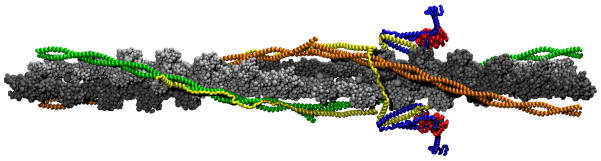
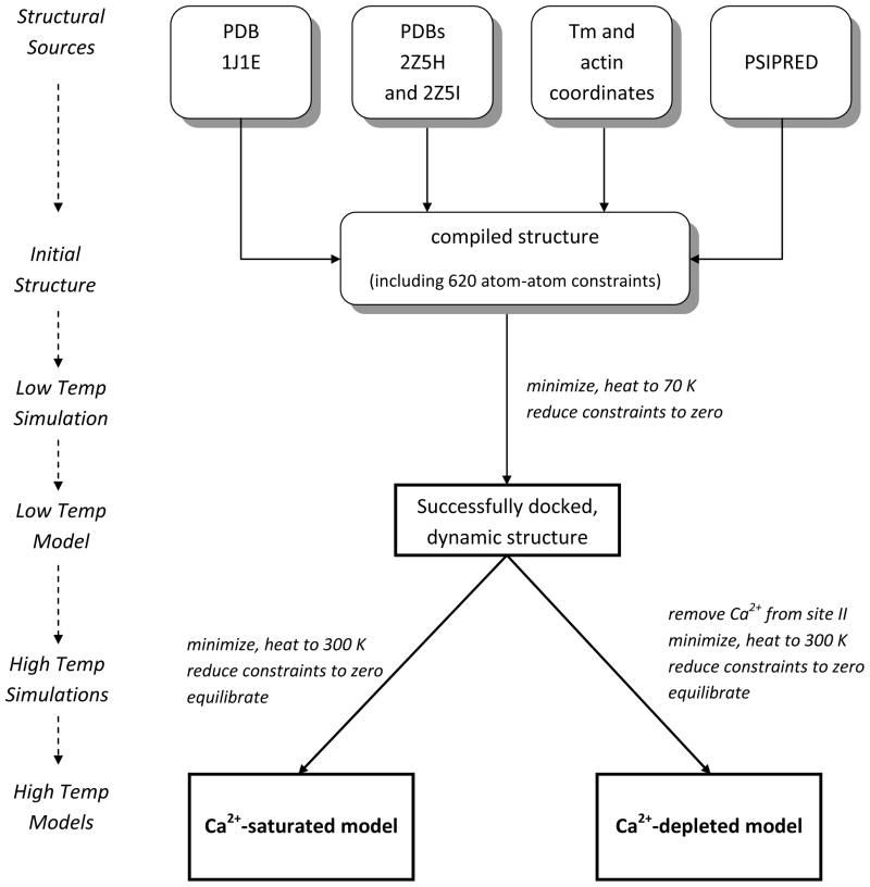
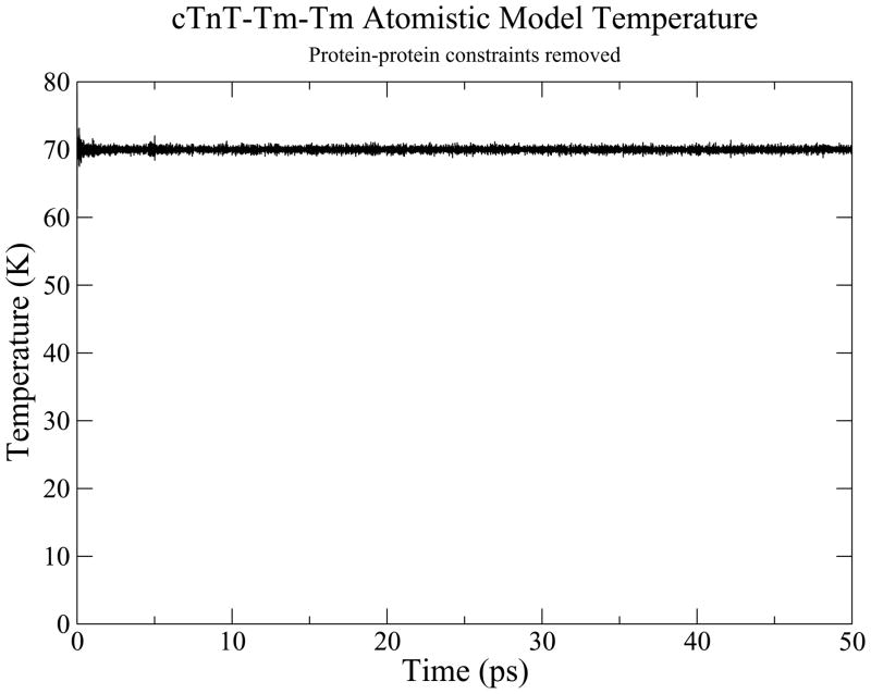
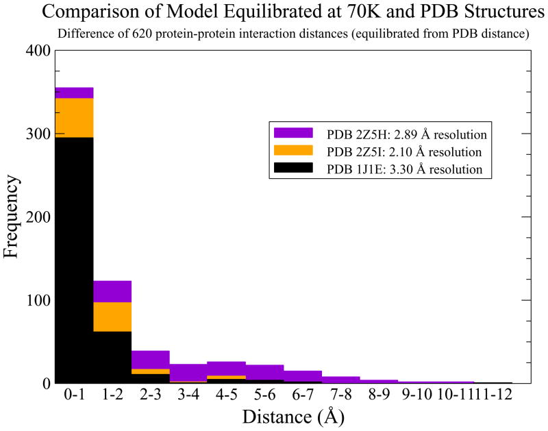
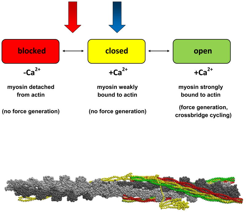
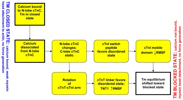
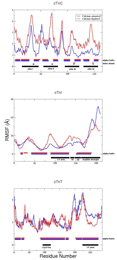
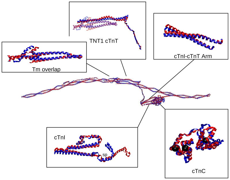
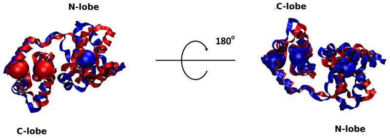
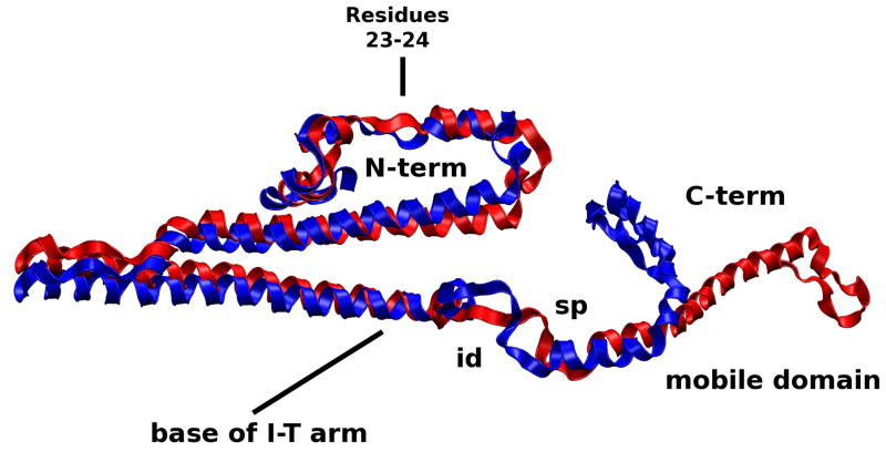
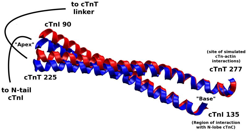
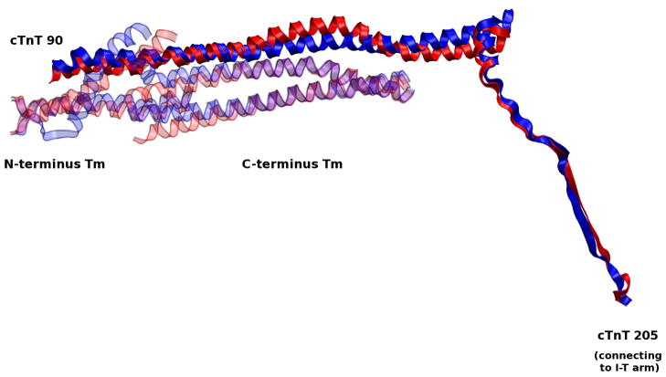
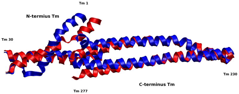
The cardiac thin filament regulates actomyosin interactions through calcium-dependent alterations in the dynamics of cardiac troponin and tropomyosin. Over the past several decades, many details of the structure and function of the cardiac thin filament and its components have been elucidated. We propose a dynamic, complete model of the thin filament that encompasses known structures of cardiac troponin, tropomyosin, and actin and show that it is able to capture key experimental findings. By performing molecular dynamics simulations under two conditions, one with calcium bound and the other without calcium bound to site II of cardiac troponin C (cTnC), we found that subtle changes in structure and protein contacts within cardiac troponin resulted in sweeping changes throughout the complex that alter tropomyosin (Tm) dynamics and cardiac troponin--actin interactions. Significant calcium-dependent changes in dynamics occur throughout the cardiac troponin complex, resulting from the combination of the following: structural changes in the N-lobe of cTnC at and adjacent to sites I and II and the link between them; secondary structural changes of the cardiac troponin I (cTnI) switch peptide, of the mobile domain, and in the vicinity of residue 25 of the N-terminus; secondary structural changes in the cardiac troponin T (cTnT) linker and Tm-binding regions; and small changes in cTnC-cTnI and cTnT-Tm contacts. As a result of these changes, we observe large changes in the dynamics of the following regions: the N-lobe of cTnC, the mobile domain of cTnI, the I-T arm, the cTnT linker, and overlapping Tm. Our model demonstrates a comprehensive mechanism for calcium activation of the cardiac thin filament consistent with previous, independent experimental findings. This model provides a valuable tool for research into the normal physiology of cardiac myofilaments and a template for studying cardiac thin filament mutations that cause human cardiomyopathies.


Figures

Figure 1
Figure 1
Representation of the human thin filament containing human cTn, Tm and actin: yellow=cTnT; blue=cTnI; red=cTnC; cyan=calcium ion; green/orange=overlapping Tm; silver/gray=actin filament (The same color scheme is used in the provided movies, except calcium ions in the movie are colored black vice cyan). This figure was obtained by orienting the cTn and Tm to an actin backbone, docking cTnT to overlapping Tm, and performing a 50 ps simulation in equilibrium at 70 K using CHARMM version 33b1(23) of cTn and the overlapping Tm without actin. The resulting cTn-Tm complex is shown here in duplicate with actin added.
Figure 2
Figure 2
Schematic of the building and simulation processes of our model.
Figure 3
Figure 3
Temperature monitoring for 50 ps equilibration of docked cTn-Tm complex.
Figure 4
Figure 4
Histogram of the absolute value of equilibrated atom-atom distances subtracted from PDB atom-atom distances to demonstrate the validity of the model.
Figure 5
Figure 5
Tm explores three states of equilibrium (blocked, closed, and open) while the cTn core remains relatively stationary with respect to actin,(10) This system is in constant flux between the three equilibrium positions of Tm, where an entire cardiac contraction cycle involving all three of these states is on the order of 100 ms.(15) We investigate how calcium directly affects the regulatory proteins cTn and Tm by looking at two representative equilibrium states with 1 ns simulations in the closed state and a position between the closed and blocked states and by comparing the changes between them. These two states are represented by the blue and red arrows, representing the Ca2+-saturated and Ca2+-depleted state, respectively.
Figure 6
Figure 6
A schematic of the two prong mechanism of Ca2+-activation of the thin filament via regulatory proteins. The mechanistic changes captured by our model are highlighted in yellow.
Figure 7
Figure 7
RMSF analysis for 1 ns simulations. Top: cTnC; Middle: cTnI; Bottom: cTnT. Regions of structural/functional interest are identified in black horizontally along the bottom of each graph. Average secondary structure is identified horizontally along the bottom of each graph where structured residues are blocked, blue for Ca2+-saturated and red for Ca2+-depleted, and unstructured residues are left blank. cTnI and cTnT secondary structure consists solely of α-helices (blocked) and coils (blank). “id” = inhibitory domain; “sp” = switch peptide; black circles = constraints on Cα 205 and 277 cTnT, all other atoms are allowed to move freely.
Figure 8
Figure 8
Alignment of Ca2+-saturated and Ca2+-depleted average structures of the complete cTn complex with overlapping Tm and highlighted regions of interest. The color scheme for this and the other structural images is: Blue=Ca2+-saturated; Red=Ca2+-depleted. In cTnC, the black spheres are the locations of calcium ions. In cTnI box, “sp” = switch peptide.
Figure 9
Figure 9
Alignment of Ca2+-saturated and Ca2+-depleted average structures of cTnC. Calcium ion locations are shown as large spheres colored according to their appropriate state. Note the lack of a calcium ion in the N-lobe in the Ca2+-depleted state. There is significant change in the conformation of the N-lobe while the C-lobe remains mostly unchanged. The Ca2+ ion positions bound to the C-lobe in the two states are virtually superimposed. Binding sites I and II as well as the linker between them are significantly altered as a result of removing Ca2+. This corresponds to the changes in RMSF that we see in Figure 7.
Figure 10
Figure 10
Alignment of Ca2+-saturated and Ca2+-depleted average structures of cTnI. The N-terminus and switch peptide and surrounding regions of the C-terminus interact with the N-lobe of cTnC and are altered as a function of Ca2+-binding. As a result, fluctuations in the dynamics of the N-lobe cTnC alter the dynamics of switch peptide of the C-terminus cTnI and the N-terminus of cTnI. id=inhibitory domain; sp=switch peptide.
Figure 11
Figure 11
Alignment of Ca2+-saturated and Ca2+-depleted average structures of the cTnI-cTnT coiled coil (I-T arm). This is the most stable region of the entire complex (in terms of subunit-subunit interactions), allowing the I-T arm to behave as united pair. The stable portion of the arm (in terms of dynamics) is at the “base” of the arm which is where cTnI interacts with the C-lobe of cTnC and where cTnT is simulated to interact with actin. The stable base allows the “apex” of the I-T arm to rotate due to fluctuations in the N-terminus of cTnI. The I-T arm rotation causes fluctuations in the cTnT linker.
Figure 12
Figure 12
Alignment of Ca2+-saturated and Ca2+-depleted average structures of the cTnT linker. Fluctuations from the apex of the I-T arm are passed on through the cTnT linker to overlapping Tm.
Figure 13
Figure 13
Alignment of Ca2+-saturated and Ca2+-depleted average structures of overlapping Tm. The overlapping region of Tm is important for its affinity to actin and flexibility.(15) The significant changes we observe in this region appear despite the fact that both strands of Tm are constrained at each end by the remaining Tm in the closed position. The loss of secondary structure we observe in these average structures is likely the result of resistance to fluctuations by these constraints, an indicator that changes in this region could shift the equilibrium of more than just the overlap region. We expect that the loss of secondary structure would not occur if the remainder of Tm was allowed to move freely and that Tm would shift toward the closed state when our model is in the Ca2+-depleted state, and possibly toward the open state when our model is in the Ca2+-saturated state which would work in tandem with actomyosin interactions. In our model residues 245–277 of the C-terminus Tm and residues 1–30 of the N-terminus Tm are allowed to move freely, the remaining residues have their alpha carbons fixed in the closed position.

References

    1. Ertz-Berger B, He H, Dowell C, Factor S, Haim T, Nunez S, Schwartz SD, Ing-wall J, Tardiff J. Changes in the chemical and dynamic properties of cardiac troponin T cause discrete cardiomyopathies in transgenic mice. Proc Natl Acad Sci USA. 2005;102:18219–18224. - PMC - PubMed
    1. Guinto PJ, Manning EP, Schwartz SD, Tardiff JC. Computational characterization of mutations in cardiac troponin T known to cause familial hypertrophic cardiomyopathy. J Theor Comp Chem. 2007;6:413–419. - PMC - PubMed
    1. Kobayashi T, Solaro RJ. Calcium, Thin Filaments, and the Integrative Biology of Cardiac Contractility. Annu Rev Physiol. 2005;67:39–67. - PubMed
    1. Tobacman LS. Thin filament-mediated regulation of cardiac contraction. Ann Rev Physiol. 1996;58:447–481. - PubMed
    1. Galinska A, Hatch V, Craig R, Murphy AM, Eyk JEV, Wang CLA, Lehman W, Foster DB. The C Terminus of Cardiac Troponin I Stabilizes the Ca(2+)-Activated State of Tropomyosin on Actin Filaments. Circ Res. 2010;106:705–711. - PMC - PubMed

Publication types