

doi: 10.1103/PhysRevLett.107.028101.
Epub 2011 Jul 8.
Cell surface as a fractal: normal and cancerous cervical cells demonstrate different fractal behavior of surface adhesion maps at the nanoscale
Affiliations
- PMID: 21797643
- PMCID: PMC5562016
- DOI: 10.1103/PhysRevLett.107.028101
Item in Clipboard
Cell surface as a fractal: normal and cancerous cervical cells demonstrate different fractal behavior of surface adhesion maps at the nanoscale
Phys Rev Lett.
.
Abstract
Here we show that the surface of human cervical epithelial cells demonstrates substantially different fractal behavior when the cell becomes cancerous. Analyzing the adhesion maps of individual cervical cells, which were obtained using the atomic force microscopy operating in the HarmoniX mode, we found that cancerous cells demonstrate simple fractal behavior, whereas normal cells can only be approximated at best as multifractal. Tested on ~300 cells collected from 12 humans, the fractal dimensionality of cancerous cells is found to be unambiguously higher than that for normal cells.
Figures

(color online). Representative AFM HarmoniX images and Fourier spectra of cancer and normal cells. Panels (a), (b) show topographical images whereas (e),(f) demonstrate the adhesion maps of cell surface. The corresponding Fourier spectra (the magnitude) as a function of reciprocal space are shown in (c),(d) and (g),(h) for topography and adhesion, respectively. The fractal behavior corresponds to straight (shown dashed) lines. The left (right) panels show the data for cancerous (normal) cells.

(color online). Distributions of fractal dimensionality (FD) for adhesion maps of cancer and normal cells calculated on the range of features L ~ 40–300 nm size. Each value shown corresponds to a single cell, which was found as an average of the fractal dimensionalities calculated on several square micron maps collected over 4 areas on each cell (the variation of the FD between the areas was less than 3% over each cell).
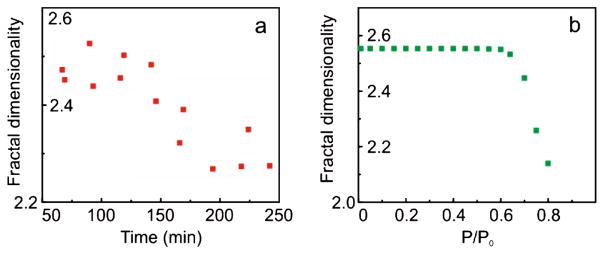
(color online). (a) An example of the dependence of fractal dimensionality (FD) of cancer cell surface maps of adhesion on time lapsed after extracting the cells from a desiccator. The imaging was done in humidity of ~70%. (b) Modeling of FD as a function of humidity.
References
-
- Mandelbrot BB. Science. 1998;279:783c.
-
- Meakin P. Fractals, Scaling, and Growth Far from Equilibrium. Cambridge University Press; Cambridge, England: 1998. p. xiv.
-
- McCauley JL. Chaos, Dynamics, and Fractals: An Algorithmic Approach to Deterministic Chaos. Cambridge University Press; Cambridge, England: 1993. p. xxi.
-
- Wu KKS, Lahav O, Rees MJ. Nature (London) 1999;397:225.
-
- Burrough PA. Nature (London) 1981;294:240.
Publication types
MeSH terms
Grants and funding
LinkOut - more resources
Full Text Sources
Other Literature Sources
Medical