

Hyaluronidase and collagenase increase the transfection efficiency of gene electrotransfer in various murine tumors
- PMID: 21797718
- PMCID: PMC3260441
- DOI: 10.1089/hum.2011.073
Hyaluronidase and collagenase increase the transfection efficiency of gene electrotransfer in various murine tumors
Abstract
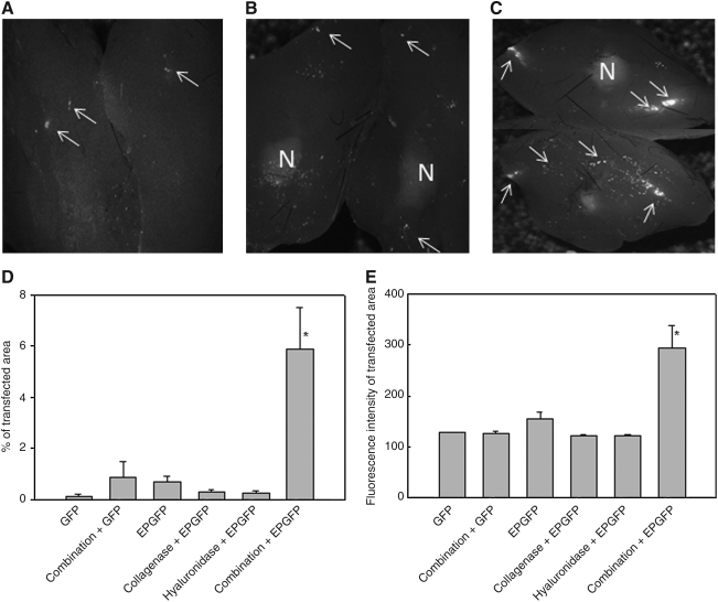
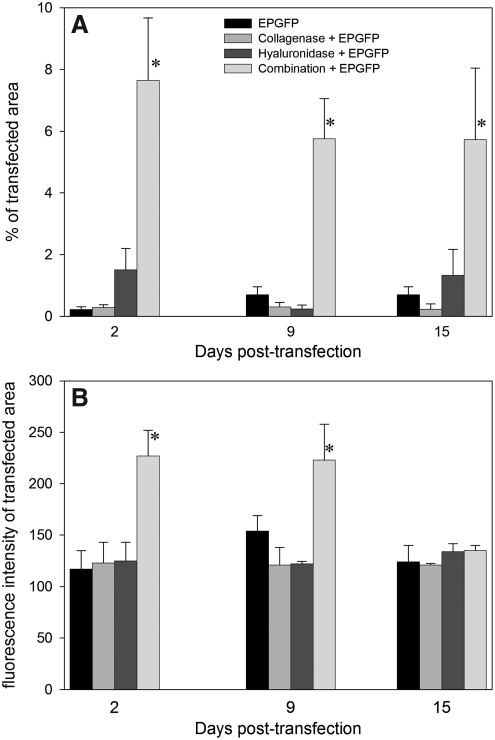
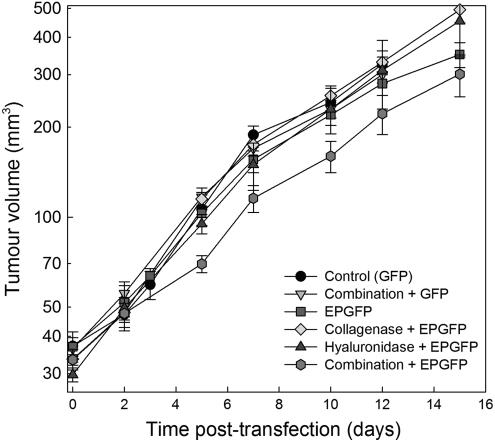
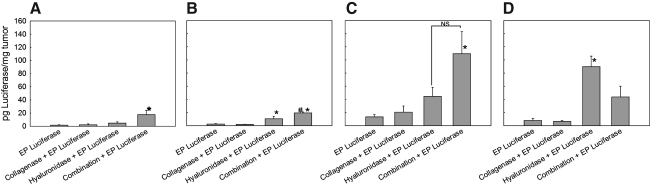
One of the applications of electroporation/electropulsation in biomedicine is gene electrotransfer, the wider use of which is hindered by low transfection efficiency in vivo compared with viral vectors. The aim of our study was to determine whether modulation of the extracellular matrix in solid tumors, using collagenase and hyaluronidase, could increase the transfection efficiency of gene electrotransfer in histologically different solid subcutaneous tumors in mice. Tumors were treated with enzymes before electrotransfer of plasmid DNA encoding either green fluorescent protein or luciferase. Transfection efficiency was determined 3, 9, and 15 days posttransfection. We demonstrated that pretreatment of tumors with a combination of enzymes significantly increased the transfection efficiency of electrotransfer in tumors with a high extracellular matrix area (LPB fibrosarcoma). In tumors with a smaller extracellular matrix area and less organized collagen lattice, the increase was not so pronounced (SA-1 fibrosarcoma and EAT carcinoma), whereas in B16 melanoma, in which only traces of collagen are present, pretreatment of tumors with hyaluronidase alone was more efficient than pretreatment with both enzymes. In conclusion, our results suggest that modification of the extracellular matrix could improve distribution of plasmid DNA in solid subcutaneous tumors, demonstrated by an increase in transfection efficiency, and thus have important clinical implications for electrogene therapy.
Figures

Similar articles
-
The effect of the histological properties of tumors on transfection efficiency of electrically assisted gene delivery to solid tumors in mice.Gene Ther. 2007 Sep;14(17):1261-9. doi: 10.1038/sj.gt.3302989. Epub 2007 Jun 28. Gene Ther. 2007. PMID: 17597791
-
Control by pulse parameters of DNA electrotransfer into solid tumors in mice.Gene Ther. 2009 May;16(5):635-44. doi: 10.1038/gt.2009.10. Epub 2009 Feb 12. Gene Ther. 2009. PMID: 19212425
-
Sequence and time dependence of transfection efficiency of electrically-assisted gene delivery to tumors in mice.Curr Drug Deliv. 2006 Jan;3(1):77-81. doi: 10.2174/156720106775197556. Curr Drug Deliv. 2006. PMID: 16472096
-
A Critical Review of Electroporation as A Plasmid Delivery System in Mouse Skeletal Muscle.Int J Mol Sci. 2019 Jun 6;20(11):2776. doi: 10.3390/ijms20112776. Int J Mol Sci. 2019. PMID: 31174257 Free PMC article. Review.
-
Electrically-assisted nucleic acids delivery to tissues in vivo: where do we stand?Curr Pharm Des. 2006;12(29):3817-25. doi: 10.2174/138161206778559740. Curr Pharm Des. 2006. PMID: 17073680 Review.
Cited by
-
Targeting tumor extracellular matrix activates the tumor-draining lymph nodes.Cancer Immunol Immunother. 2022 Dec;71(12):2957-2968. doi: 10.1007/s00262-022-03212-6. Epub 2022 May 7. Cancer Immunol Immunother. 2022. PMID: 35524791 Free PMC article.
-
In Vitro and in vivo Evaluation of Electrochemotherapy with trans-platinum Analogue trans-[PtCl2(3-Hmpy)2].Radiol Oncol. 2017 Sep 14;51(3):295-306. doi: 10.1515/raon-2017-0034. eCollection 2017 Sep. Radiol Oncol. 2017. PMID: 28959166 Free PMC article.
-
Gene Immunotherapy of Colon Carcinoma with IL-2 and IL-12 Using Gene Electrotransfer.Int J Mol Sci. 2023 Aug 17;24(16):12900. doi: 10.3390/ijms241612900. Int J Mol Sci. 2023. PMID: 37629081 Free PMC article.
-
Intratumoral electroporation of a self-amplifying RNA expressing IL-12 induces antitumor effects in mouse models of cancer.Mol Ther Nucleic Acids. 2022 Jul 20;29:387-399. doi: 10.1016/j.omtn.2022.07.020. eCollection 2022 Sep 13. Mol Ther Nucleic Acids. 2022. PMID: 36035753 Free PMC article.
-
In Vivo Evaluation of a New Recombinant Hyaluronidase to Improve Gene Electro-Transfer Protocols for DNA-Based Drug Delivery against Cancer.Cancers (Basel). 2018 Oct 28;10(11):405. doi: 10.3390/cancers10110405. Cancers (Basel). 2018. PMID: 30373297 Free PMC article.
References
-
- Aihara H. Miyazaki J. Gene transfer into muscle by electroporation in vivo. Nat. Biotechnol. 1998;16:867–870. - PubMed
-
- Alexandrakis G. Brown E.B. Tong R.T., et al. Two-photon fluorescence correlation microscopy reveals the two-phase nature of transport in tumors. Nat. Med. 2004;10:203–207. - PubMed
-
- Bancroft J.D. Cook H.C. Manual of Histological Techniques and Their Diagnostic Application. Churchill Livingstone; New York: 1994.
-
- Baumgartner G. Gomar-Hoss C. Sakr L., et al. The impact of extracellular matrix on the chemoresistance of solid tumors: Experimental and clinical results of hyaluronidase as additive to cytostatic chemotherapy. Cancer Lett. 1998;131:85–99. - PubMed
-
- Bettan M. Ivanov M.A. Mir L.M., et al. Efficient DNA electrotransfer into tumors. Bioelectrochemistry. 2000;52:83–90. - PubMed
Publication types
MeSH terms
Substances
LinkOut - more resources
Full Text Sources
Other Literature Sources
