. 2011 Jul 7:1:25.
doi: 10.1186/2044-5040-1-25.

Differentiation and fiber type-specific activity of a muscle creatine kinase intronic enhancer

Affiliations

Differentiation and fiber type-specific activity of a muscle creatine kinase intronic enhancer

Phillip Wl Tai et al. Skelet Muscle. .

Abstract

Background: Hundreds of genes, including muscle creatine kinase (MCK), are differentially expressed in fast- and slow-twitch muscle fibers, but the fiber type-specific regulatory mechanisms are not well understood.

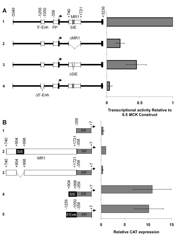
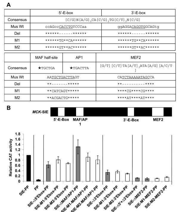
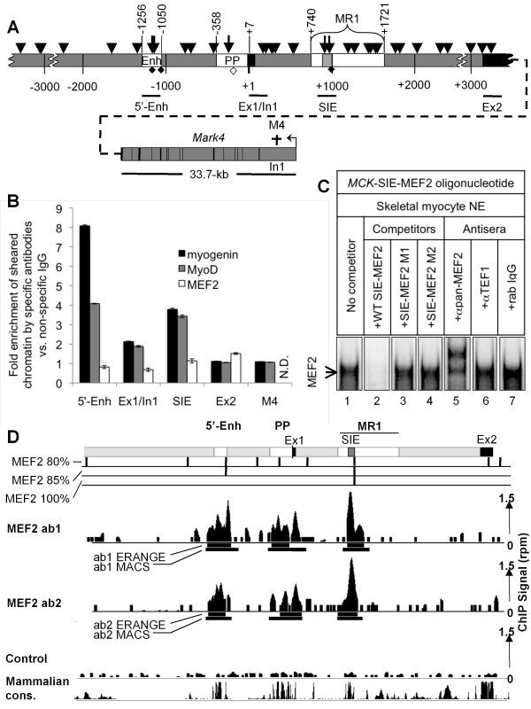
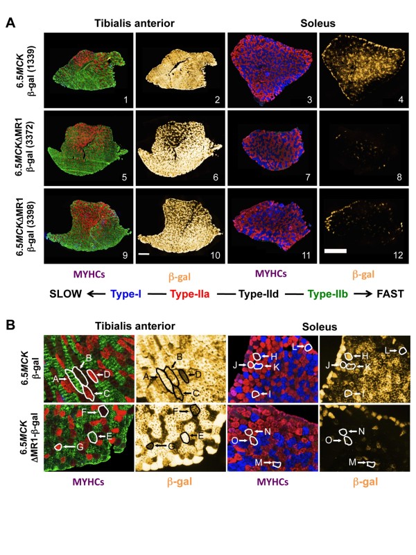
Results: Modulatory region 1 (MR1) is a 1-kb regulatory region within MCK intron 1 that is highly active in terminally differentiating skeletal myocytes in vitro. A MCK small intronic enhancer (MCK-SIE) containing a paired E-box/myocyte enhancer factor 2 (MEF2) regulatory motif resides within MR1. The SIE's transcriptional activity equals that of the extensively characterized 206-bp MCK 5'-enhancer, but the MCK-SIE is flanked by regions that can repress its activity via the individual and combined effects of about 15 different but highly conserved 9- to 24-bp sequences. ChIP and ChIP-Seq analyses indicate that the SIE and the MCK 5'-enhancer are occupied by MyoD, myogenin and MEF2. Many other E-boxes located within or immediately adjacent to intron 1 are not occupied by MyoD or myogenin. Transgenic analysis of a 6.5-kb MCK genomic fragment containing the 5'-enhancer and proximal promoter plus the 3.2-kb intron 1, with and without MR1, indicates that MR1 is critical for MCK expression in slow- and intermediate-twitch muscle fibers (types I and IIa, respectively), but is not required for expression in fast-twitch muscle fibers (types IIb and IId).

Conclusions: In this study, we discovered that MR1 is critical for MCK expression in slow- and intermediate-twitch muscle fibers and that MR1's positive transcriptional activity depends on a paired E-box MEF2 site motif within a SIE. This is the first study to delineate the DNA controls for MCK expression in different skeletal muscle fiber types.


Figures

Figure 1
Figure 1
Modulatory region 1 (MR1) contains a highly conserved subregion containing known myogenic control element motifs. Sequence alignment of MR1 reveals a highly conserved 95-bp subregion, muscle creatine kinase (MCK) small intronic enhancer (MCK-SIE), that contains five putative control elements: an E-box motif pair, a myocyte enhancer factor 2 (MEF2) consensus motif and partially overlapping sequences that match proven MAF half-site and activator protein 1 (AP-1) sequences (see also Additional file 1 Figure S1). Bases that are identical in all six species (Homo sapiens, Felis catus, Bos taurus, Sus scrota, Canis familiaris and Mus musculus) are shown in black, while bases conserved between at least three species are shown in gray. The 3'-E-box is present in all six species, but is slightly more 5' in the mouse and further 5' in the dog. Conformation of mouse control element sequences to the MyoD/myogenin and MEF2 consensus sequences are indicated below the mouse sequence (+ = conforms, - = differs).
Figure 2
Figure 2
MR1 is a positive regulator of MCK transcription. (A) MM14 skeletal myocytes were cotransfected with an MCK enhancer-alkaline phosphatase (AP) reference plasmid and test gene plasmids containing the chloramphenicol acetyl transferase (CAT) reporter gene driven by the full-length 6.5-kb MCK construct (6.5MCK-CAT, #1), the 6.5-kb construct with MR1 deleted (6.5MCKΔMR1-CAT, #2), the 6.5-kb construct with the MCK-SIE deleted (6.5MCKΔSIE-CAT, #3) or, for comparison, the 6.5-kb construct with the 5'-enhancer deleted (6.5MCKΔEnh-CAT, #4). Test construct activities are represented as the average values of relative CAT over AP activity normalized to the activity of 6.5MCK-CAT. (B) MR1 is composed of regions that promote transcription in MM14 cultures. Constructs containing the "full-length" MR1 (MR1-PP-CAT, #2), a construct lacking the MCK-SIE (MR1ΔSIE-PP-CAT, #3) or just the MCK-SIE (SIE-PP-CAT, #4) were generated to test the functional activity of the MCK-SIE. Activities of these test constructs were normalized to activities of the proximal promoter alone (PP-CAT, #1). The activity of the 5'-enhancer (5'Enh-PP-CAT, #5) is provided for comparison. Each experiment was performed in at least twelve plates in three separate experiments, and activities are averages of those experiments. Error bars represent ±1 standard deviation.
Figure 3
Figure 3
Two E-boxes and a MEF2 site are critical for activity of the MCK-SIE. (A) Deletions and mutations tested in MCK-SIE. The currently accepted consensus motifs for the E-box and MEF2 motifs are shown. Proven MAF half-site and AP-1 control element sequences are also indicated. Stars indicate sequences that were experimentally proven to recruit the labeled factors and do not represent consensus binding motifs. The wild-type mouse sequences of these elements within the MCK-SIE (Wt), the deletion sequences (Del) and two mutation sequences (M1 and M2) used in this study are shown on successive lines. Base pair deletions are indicated as hyphens, point mutations are shown as changed bases and asterisks indicate unchanged bases. (B) Mutational analysis of control elements within the MCK-SIE. The E-box, MAF/AP-1 and MEF2 motifs in the MCK-proximal promoter-CAT (MCK-SIE-PP-CAT) (diagrammed with elements in their relative positions) were deleted (gray bars) or subjected to two mutations (white bars) within core bases (Figure 2A) and were tested for transcriptional activity in differentiated MM14 skeletal myocyte cultures. The relative activities of these constructs were compared to the MCK-SIE-PP-CAT construct (scaled to equal 1.0) and PP-CAT alone (black bars). Each construct was tested in twelve plates in three separate experiments, and activities shown are averages of those experiments. Error bars represent ±1 standard deviation.
Figure 4
Figure 4
MyoD and myogenin are enriched at the MCK-SIE in skeletal myocytes. (A) Diagram of the 6.5-kb MCK regulatory region with the three known active regulatory regions: the 5'-enhancer, PP, MR1 (white boxes), the MCK-SIE (light gray box) exons 1 and 2 (black boxes) and other regions (gray), including the 33.7-kb Mark4 gene (located approximately 40 kb 3' of the MCK-SIE and transcribed in the opposite direction). E-box CAnnTG core motifs (arrowheads) occur throughout the 6.5-kb sequence. Among the thirty-five total E-boxes are two functional E-boxes within the 5'-enhancer, one functional E-box within the proximal promoter and two E-box motifs within the MCK-SIE (longer arrows). The less frequent MEF2 motifs (full diamonds) are found only in the 5'-enhancer and MCK-SIE and as a possible nonconsensus MEF2 site (open diamond) in the proximal promoter. The chromatin immunoprecipitation (ChIP) primer pairs (black lines) that span the 5'-enhancer sequence were used as positive controls for MyoD and myogenin binding to functional E-boxes. Negative controls consist of genomic regions containing either no core E-box motifs (region within the Mark4 intron 1 (M4, dagger)) or core E-box motifs with no proven transcriptional function (MCK gene exon 1/intron 1 boundary (two E-boxes) and exon 2 (one E-box); see Results, section-5). (B) MyoD and myogenin bind MCK gene E-box motifs. ChIP analyses using antibodies for MyoD, myogenin, MEF2 and control immunoglobulin G (IgG) were performed using chromatin from differentiated MM14 cell myocytes. The graph shows data from one of three ChIP experiments that is representative of the enrichment detected at each position by antibodies to myogenin (black bars), MyoD (gray bars) or MEF2 (white bars) over nonspecific rabbit IgG as determined by quantitative polymerase chain reaction (qPCR) assay. Error bars represent ±1 standard deviation of triplicate samples. (C) Electrophoretic mobility shift assay (EMSA) of MEF2 binding to the MCK-SIE MEF2 control element. Nuclear extracts from differentiated MM14 cultures were incubated with a 32P-labeled probe containing the MCK-SIE-MEF2 sequence with no competitor (lane 1), wild-type MEF2 competitor (lane 2), two different mutant MEF2 competitors (lanes 3 and 4), pan-MEF2 antibodies (lane 5), transcriptional enhancer factor 1 (TEF-1)-specific antibodies (lane 6) or nonspecific rabbit IgG (lane 7). Arrows indicate the MEF2-containing complex and free probe. (D) MEF2 ChIP-Seq occupancy at the 6.5-kb MCK regulatory region in differentiated C2 C12 cells shows that MEF2 is present at all three control regions. The 6.5-kb region is shown in schematic at the top (5'-enhancer, proximal promoter and MR1 are shown in white; MCK-SIE is shown in gray). Sequences that match the MEF2 canonical motif (CTAWWWWTAG) at the 80%, 85% and 100% thresholds are mapped throughout the 6.5-kb region. The sequenced and mapped ChIP signals (reads per million (rpm)) for the two pan-MEF2 antibodies 1 and 2 and the control (input DNA) are indicated as black histograms (scale shown at the right). Two different ChIP-Seq region finders (Model-based Analysis of ChIP-Seq data and Enhanced Read Analysis of Gene Expression) define the sequence range in which MEF2 is predicted to bind (see Materials and methods), and these are shown below each signal track as black bars. Conservation across the regions is shown from the University of California Santa Cruz (UCSC) Genome Browser plot of phastCons scores for the 20 default placental mammals.
Figure 5
Figure 5
MR1 is important for MCK expression in slow- and intermediate-twitch skeletal muscle fibers. (A) Sister sections of tibialis anterior (TA) and soleus muscles from mice carrying the 6.5MCK-β-gal or the 6.5ΔMR1-β-gal transgenes, immunostained with myosin heavy chain (MYHC) fiber type-specific monoclonal antibodies (panels 1, 3, 5, 7, 9 and 11) or activity stained for β-galactosidase (β-gal) expression (panels 2, 4, 6, 8, 10 and 12). Antibodies for different isoforms and fluorophore-labeled secondary antibodies mark the fiber types as follows: slow-twitch fibers (type I), blue; intermediate-twitch fibers (type IIa), red; and fast-twitch fibers (types IIb and IId), green and black, respectively (the black appearance of type IId fibers is due to the absence of any type 1, IIa, or IIb antibody binding). Purplish fibers contain both types I and IIa MYHCs (see Figure 5B, soleus), and fibers with weak red or green staining probably contain mixtures of type IId (no color) + type IIa or type IId + type IIb, respectively (see Figure 5B, TA). Sister sections were stained for β-gal expression (false colored gold). Bars are 0.5 mm. (B) Higher magnification sections indicate differences in β-gal expression between fiber types in transgenic lines with and without MR1. Individual fibers, outlined in white or black to show relative differences in X-gal staining between fiber types (type I = K, L and O; type IIa = C, D, G, I and J; type IId = B, F, H and M; and type IIb = A and E), can be cross-referenced to β-gal expression in sister sections.

Similar articles

Cited by

References

    1. Welle S, Bhatt K, Thornton CA. Inventory of high-abundance mRNAs in skeletal muscle of normal men. Genome Res. 1999;9:506–513. - PMC - PubMed
    1. Chamberlain JS, Jaynes JB, Hauschka SD. Regulation of creatine kinase induction in differentiating mouse myoblasts. Mol Cell Biol. 1985;5:484–492. - PMC - PubMed
    1. Tapscott SJ. The circuitry of a master switch: Myod and the regulation of skeletal muscle gene transcription. Development. 2005;132:2685–2695. doi: 10.1242/dev.01874. - DOI - PubMed
    1. Lyons GE, Muhlebach S, Moser A, Masood R, Paterson BM, Buckingham ME, Perriard JC. Developmental regulation of creatine kinase gene expression by myogenic factors in embryonic mouse and chick skeletal muscle. Development. 1991;113:1017–1029. - PubMed
    1. Yamashita K, Yoshioka T. Profiles of creatine kinase isoenzyme compositions in single muscle fibres of different types. J Muscle Res Cell Motil. 1991;12:37–44. doi: 10.1007/BF01781172. - DOI - PubMed

LinkOut - more resources