. 2011 Jul 13;1(1):26.
doi: 10.1186/2044-5040-1-26.

T-tubule biogenesis and triad formation in skeletal muscle and implication in human diseases

Affiliations

T-tubule biogenesis and triad formation in skeletal muscle and implication in human diseases

Lama Al-Qusairi et al. Skelet Muscle. .

Abstract

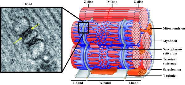
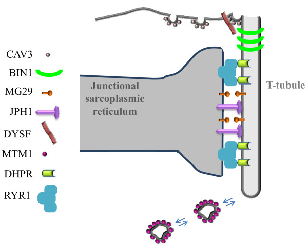
In skeletal muscle, the excitation-contraction (EC) coupling machinery mediates the translation of the action potential transmitted by the nerve into intracellular calcium release and muscle contraction. EC coupling requires a highly specialized membranous structure, the triad, composed of a central T-tubule surrounded by two terminal cisternae from the sarcoplasmic reticulum. While several proteins located on these structures have been identified, mechanisms governing T-tubule biogenesis and triad formation remain largely unknown. Here, we provide a description of triad structure and plasticity and review the role of proteins that have been linked to T-tubule biogenesis and triad formation and/or maintenance specifically in skeletal muscle: caveolin 3, amphiphysin 2, dysferlin, mitsugumins, junctophilins, myotubularin, ryanodine receptor, and dihydhropyridine Receptor. The importance of these proteins in triad biogenesis and subsequently in muscle contraction is sustained by studies on animal models and by the direct implication of most of these proteins in human myopathies.


Figures

Figure 1
Figure 1
Triad organization in skeletal muscle. Left: Electron micrograph of a triad junction. A central T-tubule is flanked on both sides by a terminal cisternae element from the sarcoplasmic reticulum. Arrows indicate electron-dense junctional feet corresponding to the ryanodine receptor-dihydhropyridine receptor complex. Right: Schematic representation of a mammalian muscle sarcomere and surrounding membranes. T-tubules shown in gray are specialized invaginations of the sarcolemma. The elaborated sarcoplasmic reticulum network is shown in blue. Note the close proximity of T-tubules and terminal cisternae of the sarcoplasmic reticulum (adapted from [104]; © 2007 by Pearson Education, Inc.).
Figure 2
Figure 2
Dynamics of T-tubule vacuolation produced by the efflux of glycerol. A single frog skeletal muscle fiber stained with a lipophilic fluorescence probe is shown. Shown are serial confocal microscopic images of the same fiber (a) 5 minutes, (b) 12 minutes and (c) 30 minutes after the fiber was transferred from a solution containing 110 mM glycerol to a solution without glycerol (reprinted from [10]; © 2001 with permission from Elsevier).
Figure 3
Figure 3
Proteins implicated in triad organization in skeletal muscle. (a) Electron microscopic image showing ferrocyanate-treated muscle from wild-type mouse. (b) Electron microscopic image showing ferrocyanate-treated muscle from Cav3 (caveolin 3)-mutant mouse. Note the altered appearance and orientation of T-tubules (from [32]; © 2001 The American Society for Biochemistry and Molecular Biology). (c) Electron microscopic image from Drosophila normal muscle. Note that dyads are localized in proximity to the Z-line (similar to vertebrate). (d through f) Electron microscopic images from Bin1 (amphiphysin)-null Drosophila showing alteration of T-tubules resulting in (d) mislocalized, (e) longitudinal and (f) dilated tubules (from [43]; © 2001 Cold Spring Harbor Laboatory Press). (g through j) Electron microscopic images of ferrocyanate-treated muscles from (g and h) wild-type and (i and j) MG29 (mitsugumin)-knockout mice demonstrating the alterations in T-tubule organization. Note the absence of (i) T-tubule or (j) longitudinal orientation (from [61]; © 1999 The Rockefeller University Press. The Journal of Cell Biology, 2002, 159:695-705). (k and l) Electron microscopic images from (k) wild-type and (l) JPH1 (junctophilin)-knockout skeletal muscles from neonate mice showing the altered triads with swollen SR (reprinted from [73]; © 2002 with permission from Elsevier). (m and n) Electron microscopic images of ferrocyanate-treated muscles from (m) wild-type and (n) Dysf (dysferlin)-knockout mice showing an abnormal shape of the T-tubule system (from [51]; © 2010 John Wiley and Sons). (o and p) Electron microscopic images of ferrocyanate-treated muscles from (o) wild-type and (p) Mtm1 (myotubularin)-knockout mice revealing a severe alteration in T-tubule organization. Note the absence (arrowhead) or the longitudinally oriented (arrow) T-tubules (from [82]; © 2009 National Academy of Sciences, USA).
Figure 4
Figure 4
Hypothetical model for triad biogenesis in skeletal muscle. The different proteins discussed in the section Molecular mechanisms involved in T-tubule biogenesis and triad formation and maintenance are indicated, and their hypothetical implications in T-tubule biogenesis and triad formation and maintenance are depicted. Details can be found in the conclusion section.

References

    1. Curtis BM, Catterall WA. Purification of the calcium antagonist receptor of the voltage-sensitive calcium channel from skeletal muscle transverse tubules. Biochemistry. 1984;23:2113–2118. doi: 10.1021/bi00305a001. - DOI - PubMed
    1. Fosset M, Jaimovich E, Delpont E, Lazdunski M. [3H]nitrendipine receptors in skeletal muscle. J Biol Chem. 1983;258:6086–6092. - PubMed
    1. Inui M, Saito A, Fleischer S. Isolation of the ryanodine receptor from cardiac sarcoplasmic reticulum and identity with the feet structures. J Biol Chem. 1987;262:15637–15642. - PubMed
    1. MacLennan DH, Wong PT. Isolation of a calcium-sequestering protein from sarcoplasmic reticulum. Proc Natl Acad Sci USA. 1971;68:1231–1235. doi: 10.1073/pnas.68.6.1231. - DOI - PMC - PubMed
    1. Chen SR, Zhang L, MacLennan DH. Asymmetrical blockade of the Ca2+ release channel (ryanodine receptor) by 12-kDa FK506 binding protein. Proc Natl Acad Sci USA. 1994;91:11953–11957. doi: 10.1073/pnas.91.25.11953. - DOI - PMC - PubMed