. 2011 May 4;1(1):18.
doi: 10.1186/2044-5040-1-18.

Ryanodine receptors

Affiliations

Ryanodine receptors

E Michelle Capes et al. Skelet Muscle. .

Abstract

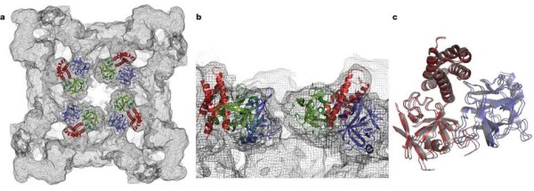
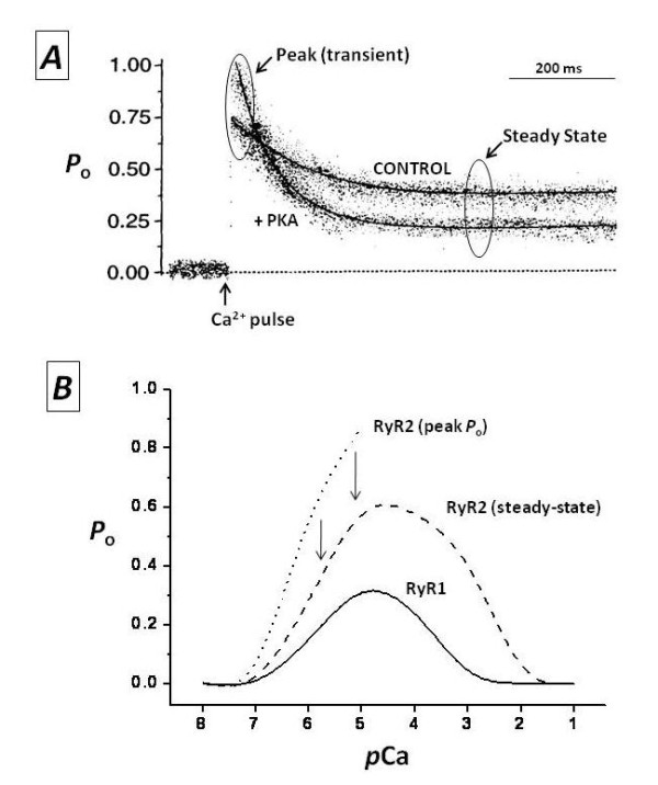
Excitation-contraction coupling involves the faithful conversion of electrical stimuli to mechanical shortening in striated muscle cells, enabled by the ubiquitous second messenger, calcium. Crucial to this process are ryanodine receptors (RyRs), the sentinels of massive intracellular calcium stores contained within the sarcoplasmic reticulum. In response to sarcolemmal depolarization, RyRs release calcium into the cytosol, facilitating mobilization of the myofilaments and enabling cell contraction. In order for the cells to relax, calcium must be rapidly resequestered or extruded from the cytosol. The sustainability of this cycle is crucially dependent upon precise regulation of RyRs by numerous cytosolic metabolites and by proteins within the lumen of the sarcoplasmic reticulum and those directly associated with the receptors in a macromolecular complex. In addition to providing the majority of the calcium necessary for contraction of cardiac and skeletal muscle, RyRs act as molecular switchboards that integrate a multitude of cytosolic signals such as dynamic and steady calcium fluctuations, β-adrenergic stimulation (phosphorylation), nitrosylation and metabolic states, and transduce these signals to the channel pore to release appropriate amounts of calcium. Indeed, dysregulation of calcium release via RyRs is associated with life-threatening diseases in both skeletal and cardiac muscle. In this paper, we briefly review some of the most outstanding structural and functional attributes of RyRs and their mechanism of regulation. Further, we address pathogenic RyR dysfunction implicated in cardiovascular disease and skeletal myopathies.


Figures

Figure 1
Figure 1
Crystallized structure of rabbit ryanodine receptor (RyR)1 for amino acid residues 1-559. This RyR1 segment folds into three distinct domains, forming a vestibule around the four-fold symmetry axis: domain A (blue; 1-205), domain B (green; 206-394) and domain C (red; 395-532). (A) Cytoplasmic view; (B) close-up lateral view from the four-fold symmetry axis, (C) Docking of domain A alone (blue) or domain BC alone (red) yields a near-perfect superposition on the docking position for ABC (grey). Adapted from Tung, et al. [17].
Figure 2
Figure 2
Modulation of ryanodine receptors (RyRs) by Ca2+. (A) Ensemble currents of single RyR2 show the effect of fast Ca2+ pulses on the Po of a non-phosphorylated channel ('control') or the same channel after phosphorylation with the catalytic subunit of protein kinase A ('+PKA'). Laser photolysis of 'caged Ca2+' (o-nitrophenyl EGTA, tetrapotassium salt; NP-EGTA) increased [Ca2+] from 0.1 to 10 μmol/l at the point labeled as 'Ca2+ pulse' (modified from [51]. (B) Ca2+-Po curves of RyR1 and RyR2. Channel activity was measured at the indicated stationary concentrations of Ca2+ ('RyR1' and 'RyR2 steady-state') and right after a fast Ca2+ pulse, as in (A) ('RyR2 (peak Po)') (unpublished results) (see text for details).

References

    1. Benitah JP, Alvarez JL, Gomez AM. L-type Ca(2+) current in ventricular cardiomyocytes. J Mol Cell Cardiol. 2010;48:26–36. doi: 10.1016/j.yjmcc.2009.07.026. - DOI - PubMed
    1. Lorenzon NM, Beam KG. Disease causing mutations of calcium channels. Channels (Austin) 2008;2:163–179. - PubMed
    1. Lanner JT, Georgiou DK, Joshi AD, Hamilton SL. Ryanodine receptors: structure, expression, molecular details, and function in calcium release. Cold Spring Harb Perspect Biol. 2010. - PMC - PubMed
    1. Coronado R, Morrissette J, Sukhareva M, Vaughan DM. Structure and function of ryanodine receptors. Am J Physiol. 1994;266:C1485–1504. - PubMed
    1. Hakamata Y, Nakai J, Takeshima H, Imoto K. Primary structure and distribution of a novel ryanodine receptor/calcium release channel from rabbit brain. FEBS Lett. 1992;312:229–235. doi: 10.1016/0014-5793(92)80941-9. - DOI - PubMed

LinkOut - more resources