. 2011 May 4;1(1):21.
doi: 10.1186/2044-5040-1-21.

Aberrant repair and fibrosis development in skeletal muscle

Affiliations

Aberrant repair and fibrosis development in skeletal muscle

Christopher J Mann et al. Skelet Muscle. .

Abstract

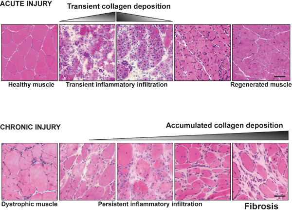
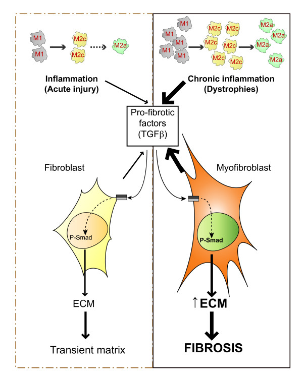
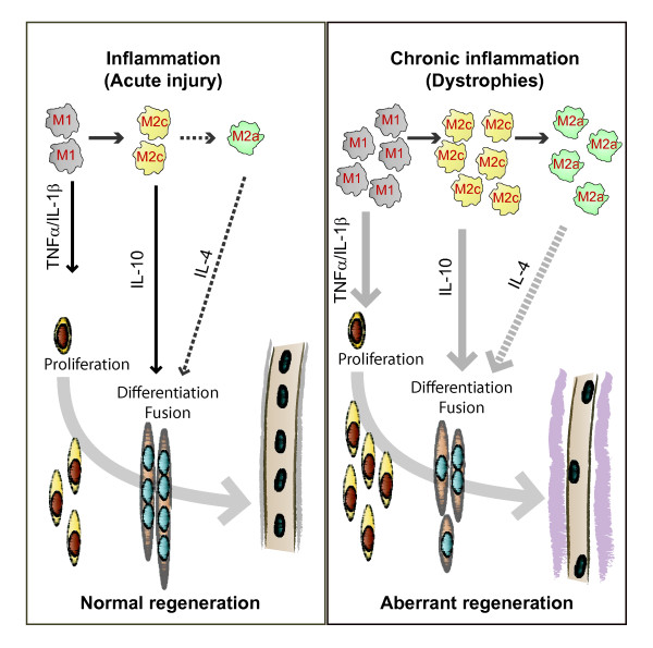
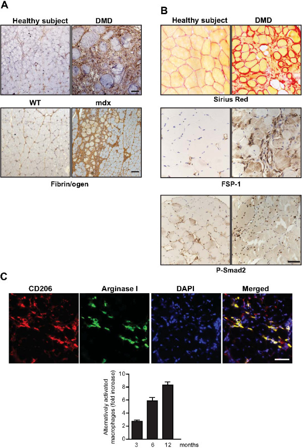
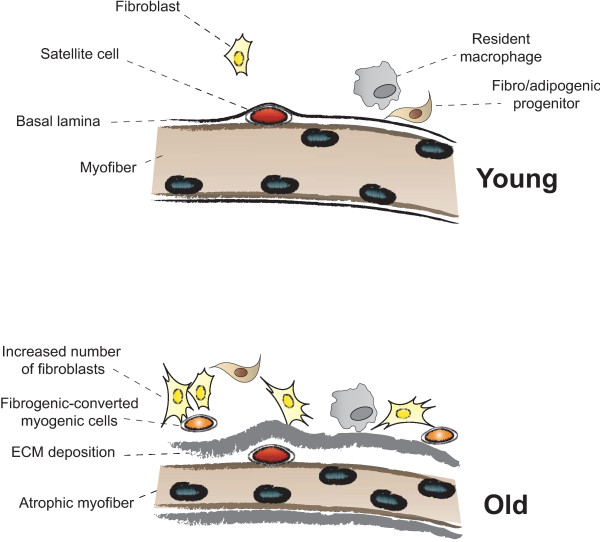
The repair process of damaged tissue involves the coordinated activities of several cell types in response to local and systemic signals. Following acute tissue injury, infiltrating inflammatory cells and resident stem cells orchestrate their activities to restore tissue homeostasis. However, during chronic tissue damage, such as in muscular dystrophies, the inflammatory-cell infiltration and fibroblast activation persists, while the reparative capacity of stem cells (satellite cells) is attenuated. Abnormal dystrophic muscle repair and its end stage, fibrosis, represent the final common pathway of virtually all chronic neurodegenerative muscular diseases. As our understanding of the pathogenesis of muscle fibrosis has progressed, it has become evident that the muscle provides a useful model for the regulation of tissue repair by the local microenvironment, showing interplay among muscle-specific stem cells, inflammatory cells, fibroblasts and extracellular matrix components of the mammalian wound-healing response. This article reviews the emerging findings of the mechanisms that underlie normal versus aberrant muscle-tissue repair.


Figures

Figure 1
Figure 1
Extracellular matrix (ECM) deposition in acute and chronic muscle regeneration. Acute injury to healthy muscle produces rapid and controlled inflammation that removes dead and damaged myofibers, and promotes replacement of the injured muscle. However, in conditions of chronic injury, as occurs in the muscular dystrophies, chronic inflammatory events result in the excessive accumulation of ECM components, which inhibit myogenic repair and lead to muscle being replaced by fibrotic/scar tissue. (Top) Tibialis anterior muscles of mice were injected with cardiotoxin and samples were taken at different stages of the regeneration process. A representative sample showing the inflammatory phase, characterized by a transient increase in collagen deposition, and subsequently the resolving phase of healing, with progressive recovery of the normal tissue morphology (hematoxylin and eosin). (Bottom) Evolution of the morphological changes seen in the diaphragm of mdx dystrophic mice with disease progression, leading to heterogeneity in fiber size and increased collagen deposition between the altered myofibers. Bars = 50 μm.
Figure 2
Figure 2
Chronic inflammation leads to fibrosis in skeletal-muscle repair. Resident and extravasating peripheral macrophages play an important role in the early stages of muscle repair after acute injury, with pro-inflammatory (M1) macrophages first acting to clear the damage, and anti-inflammatory (M2c) macrophages and alternatively activated macrophages (M2a), implicated in the subsequent resumption of inflammation, extracellular matrix (ECM) deposition and tissue repair. M2c and M2a macrophages release anti-inflammatory cytokines and pro-fibrotic molecules such as transforming growth factor (TGF)-β, which in turn activate fibroblasts in a regulated manner to produce ECM components and ECM-remodeling factors, including autocrine production of TGFβ, collagen, fibronectin, serine proteases (such as uPA/plasmin), and metalloproteinases (MMPs) and their inhibitors (TIMPs). However, during chronic tissue damage, as in muscular dystrophies, the increased and persistent presence of macrophages modify the intensity, duration and interactions of these released factors, leading to excessive ECM accumulation and replacement of muscle with fibrotic tissue.
Figure 3
Figure 3
Inflammatory control of skeletal-muscle regeneration. Replacement of damaged muscle fibers is dependent on satellite cells, resident stem cells that are normally quiescent, and are located under the basal lamina of muscle fibers. Tissue damage leads to their activation, proliferation, differentiation and fusion to form new myofibers. However, their capacity to mediate repair is modified by the extent and type of injury, and consequently by their interaction with various cellular and soluble mediators, most importantly with infiltrating macrophages. The proposed paracrine interaction between macrophages and satellite cells is as follows. During the timely, regulated process of regeneration after acute injury (left), pro-inflammatory cytokines released from M1-macrophages may promote satellite-cell proliferation, whereas cytokines released by anti-inflammatory (M2c) and alternatively activated (M2a) macrophages, respectively, may favor their differentiation and fusion. In particular, interleukin (IL)-4 was shown to regulate fusion of myoblasts in vitro and in vivo [129]. It could be expected that, during chronic damage (right), such as in muscular dystrophies, the increased and persistent presence of the distinct macrophage cell types could modify the relative levels and kinetics of these cytokines, resulting in altered satellite-cell functions and aberrant regeneration, with progressive development of fibrosis and fat accumulation, ultimately leading to non-functional muscle tissue.
Figure 4
Figure 4
Inflammatory and fibrotic traits in dystrophic muscle of patients with DMD and mdx mice. (A) Fibrin(ogen) accumulates in muscles of patients with DMD and in aged mdx mice. Immunohistochemistry for fibrin(ogen) (brown) in muscle biopsies of (top) patients with DMD and healthy subjects and in (bottom) wild-type (WT) and mdx diaphragms. (B) Increased fibrosis, fibroblast number and TGFβ signaling in dystrophic muscles. Staining for collagen deposition (Sirius red) and immunohistochemistry for fibroblast-specific protein (FSP)-1 and P-Smad2 was performed on muscle biopsies taken from patients with DMD and healthy subjects. (C) Presence of alternatively activated macrophages in diaphragm muscle of mdx mice. Cells double-positive for CD206 (red) and Arginase I (green) in mdx diaphragm are shown by immunofluorescence with specific antibodies. The relative increase in the number of these cells in the mdx diaphragm over time is shown. Bars = 50 μm.
Figure 5
Figure 5
Myofiber growth and extracellular matrix (ECM) accumulation after damage differ in young and aged muscles. (Top) In young muscles in response to injury, the satellite cells under the basal lamina can be activated by environmental cues released by the neighboring cells (a local milieu composed of fibroblasts, interstitial cells, resident macrophages, fibro/adipogenic progenitors (FAPs) and microvasculature-associated cells) to eventually form new fibers almost indistinguishable to the pre-existing ones. (Bottom) In aged muscles, the repair process will result in reduced size of newly formed myofibers and thickening of the basal lamina by enhanced deposition of ECM components, which could potentially be ascribed to the increased presence and activity of resident fibroblasts (and/or FAPs), and to decreased satellite-cell myogenic potential. Conversion of myogenic into fibrogenic cells could also contribute to ECM accumulation. These new microenvironment conditions within the satellite-cell niche will impede efficient satellite-cell functions and muscle repair.

References

    1. Emery AE. The muscular dystrophies. Lancet. 2002;359:687–695. doi: 10.1016/S0140-6736(02)07815-7. - DOI - PubMed
    1. Blake DJ, Weir A, Newey SE, Davies KE. Function and genetics of dystrophin and dystrophin-related proteins in muscle. Physiological reviews. 2002;82:291–329. - PubMed
    1. Mauro A. Satellite cells of skeletal fibers. J Biophys Biochem Cytol. 1961;9:493–495. doi: 10.1083/jcb.9.2.493. - DOI - PMC - PubMed
    1. Porter JD, Khanna S, Kaminski HJ, Rao JS, Merriam AP, Richmonds CR, Leahy P, Li J, Guo W, Andrade FH. A chronic inflammatory response dominates the skeletal muscle molecular signature in dystrophin-deficient mdx mice. Hum Mol Genet. 2002;11:263–272. doi: 10.1093/hmg/11.3.263. - DOI - PubMed
    1. Muir LA, Chamberlain JS. Emerging strategies for cell and gene therapy of the muscular dystrophies. Expert Rev Mol Med. 2009;11:e18. - PMC - PubMed