. 2011 Jul 29;333(6042):637-42.
doi: 10.1126/science.1205295.

Impaired respiratory and body temperature control upon acute serotonergic neuron inhibition

Affiliations

Impaired respiratory and body temperature control upon acute serotonergic neuron inhibition

Russell S Ray et al. Science. .

Abstract

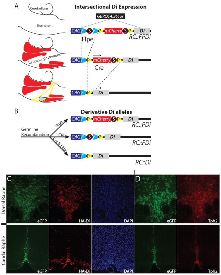
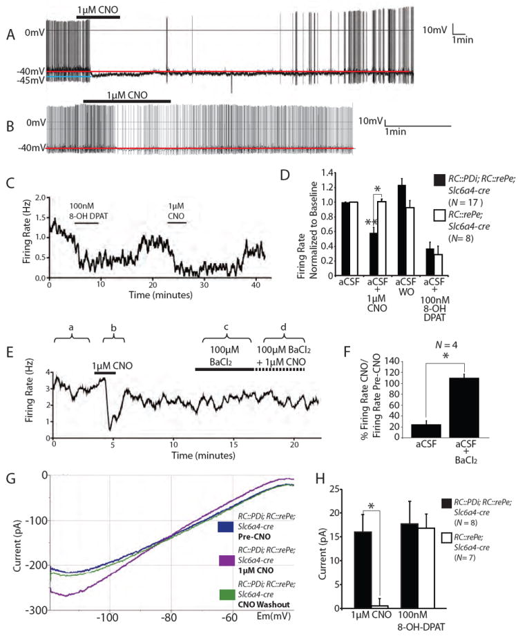
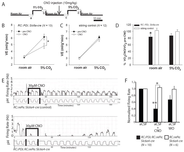
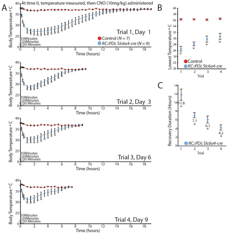
Physiological homeostasis is essential for organism survival. Highly responsive neuronal networks are involved, but their constituent neurons are just beginning to be resolved. To query brain serotonergic neurons in homeostasis, we used a neuronal silencing tool, mouse RC::FPDi (based on the synthetic G protein-coupled receptor Di), designed for cell type-specific, ligand-inducible, and reversible suppression of action potential firing. In mice harboring Di-expressing serotonergic neurons, administration of the ligand clozapine-N-oxide (CNO) by systemic injection attenuated the chemoreflex that normally increases respiration in response to tissue carbon dioxide (CO(2)) elevation and acidosis. At the cellular level, CNO suppressed firing rate increases evoked by CO(2) acidosis. Body thermoregulation at room temperature was also disrupted after CNO triggering of Di; core temperatures plummeted, then recovered. This work establishes that serotonergic neurons regulate life-sustaining respiratory and thermoregulatory networks, and demonstrates a noninvasive tool for mapping neuron function.


Conflict of interest statement

The authors declare no conflict of interests

Figures

Fig. 1
Fig. 1
Cell-selective Di expression via RC∷FPDi. (A) This Gt (ROSA)26Sor knock-in allele consists of CAG regulatory elements, an FRT-flanked transcriptional Stop, a loxP-flanked mCherry-Stop, and HA-tagged-Di-encoding sequence. A hypothetical example (left) illustrates intersectional restriction of Di to a serotonergic neuron subset (gray area, lower schema) after Flpe- and Cre-recombination; mCherry marks Flpe-only cells (red areas represent serotonergic nuclei); neither Di nor mCherry are expressed in Cre-only cells (yellow circumscribed area). (B) Derivative Di alleles. (C, D) In RC∷PDi, RC∷rePe, Slc6a4-cre brainstems, we observed concurrent reproducible expression of HA-tagged-Di (far-red indirect HA-immunofluorescence), eGFP, and Tph2 (indirect immunofluorescence). DAPI, nuclear stain 4’,6-diamidino-2-phenylindole.
Fig. 2
Fig. 2
Inducible and reversible suppression of serotonergic neuron excitability using RC∷PDi. (A, B) Recordings of cultured medullary serotonergic neurons from RC∷PDi; RC∷rePe; Slc6a4-cre mice showing CNO-induced abolishment (A) or moderate suppression (B) of action potential firing, with recovery following return to artificial cerebrospinal fluid (aCSF) superfusate. (C) Firing rate during sequential applications of 8-OH-DPAT and CNO. (D) Average firing rates (normalized to baseline ± SEM) of Di/GFP-expressing versus control neurons. Inhibition by CNO was observed for Di-neurons compared to: pre-CNO, post-CNO aCSF superfusate (aCSF washout (WO) of CNO), and to CNO-exposed control neurons, **p < 0.0001 (Friedman test), *p < 0.005 (Mann-Whitney test). CNO-inhibition was comparable to that of 8-OH-DPAT (N = 9 RC∷PDi; RC∷rePe; Slc6a4-cre neurons; N = 8 control neurons). (E) Sample trace of BaCl2-mediated block of CNO-induced suppression. (F) Average ratio of firing rate for CNO versus pre-CNO, expressed as a percentage ± SEM. Neurons were assayed in aCSF (a and b in (E)) and upon subsequent application of BaCl2 (c and d) *p < 0.05 (Friedman test). (G) Voltage clamp recording showing the current-voltage relationship of a Di-expressing serotonergic neuron with or without CNO. (H) Average current elicited by CNO and 8-OH-DPAT at -60 mV, *p = 0.002 (Mann-Whitney test).
Fig. 3
Fig. 3
CNO/Di-perturbation of serotonergic neurons disrupts the central respiratory CO2 chemoreflex. (A) Protocol for plethysmographic assessment at 34°C of respiratory responses to inspired CO2 in the awake animal before and after CNO administration; boxes (a, b, c, d) represent points analyzed from continuous recordings. (B) RC∷PDi; Slc6a4-cre mice upon CNO administration showed reduced minute ventilation responses to inspired CO2 as compared to pre-CNO baselines, *p = 0.002 (RM ANOVA followed by Tukey post-hoc). (C) Pre-CNO and CNO responses to CO2 were indistinguishable for control mice and from the pre-CNO response of RC∷PDi;Slc6a4-cre mice. (D) RC∷PDi;Slc6a4-cre mice exhibited reduced VO2 upon CNO administration as compared to controls, *p < 0.05 (paired t-test). (E) Firing rate and simultaneous bath pH recordings from cultured serotonergic neurons from controls (top) compared to RC∷PDi; RC∷rePe; Slc6a4-cre mice (bottom). Changing CO2 from 5% to 9% shifted pH from ~7.4 to ~7.16 and induced an increase in firing rate. In Di-neurons (bottom), this response was inhibited by CNO and reversed on return to aCSF. (F) Peak firing rates (normalized to baseline) in 9% CO2-saturated aCSF were suppressed with CNO application in RC∷PDi; RC∷rePe;Slc6a4-cre neurons, *p < 0.05 (Mann-Whiteney test).
Fig. 4
Fig. 4
CNO/Di-inhibition of serotonergic neurons induced severe yet reversible and repeatable hypothermia. Trials consisted of body temperature assessments taken at room temperature just before a single CNO administration and then every 10 min for the first half hour, followed by every 30 min until recovery. Animals underwent 4 sequential trials. (A) Body temperature averages of RC∷PDi; Slc6a4-cre mice versus controls before and after CNO injection, *p < 0.05 (unpaired t-test). (B) Average lowest temperatures achieved per trial, *p < 0.05 (a one-way repeated measures ANOVA). (C) Average duration to return to 36°Cfollowing CNO injection, *p < 0.01 (a one-way repeated measures ANOVA).

Comment in

References

    1. Leusen IR. Chemosensitivity of the respiratory center; influence of CO2 in the cerebral ventricles on respiration. Am J Physiol. 1954 Jan;176:39. - PubMed
    1. Kety SS, Forster RE, Comroe Julius H., Jr March 13, 1911-July 31, 1984. Biogr Mem Natl Acad Sci. 2001;79:66. - PubMed
    1. Nattie E, Comroe Julius H., Jr distinguished lecture: central chemoreception: then … and now. J Appl Physiol. 2011 Jan;110:1. - PMC - PubMed
    1. Guyenet PG, Stornetta RL, Bayliss DA. Central respiratory chemoreception. J Comp Neurol. 2010 Oct 1;518:3883. - PMC - PubMed
    1. Smith CA, Rodman JR, Chenuel BJ, Henderson KS, Dempsey JA. Response time and sensitivity of the ventilatory response to CO2 in unanesthetized intact dogs: central vs. peripheral chemoreceptors. J Appl Physiol. 2006 Jan;100:13. - PubMed

Publication types

MeSH terms