. 2011 Sep 23;286(38):33252-9.
doi: 10.1074/jbc.M111.259101. Epub 2011 Jul 28.

Rod phosphodiesterase-6 (PDE6) catalytic subunits restore cone function in a mouse model lacking cone PDE6 catalytic subunit

Affiliations

Rod phosphodiesterase-6 (PDE6) catalytic subunits restore cone function in a mouse model lacking cone PDE6 catalytic subunit

Saravanan Kolandaivelu et al. J Biol Chem. .

Abstract

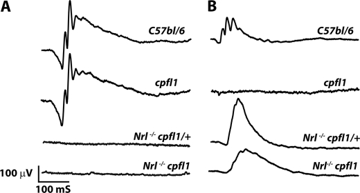
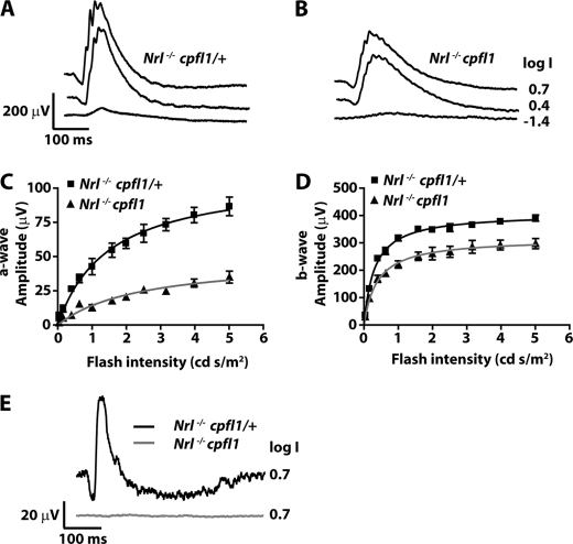
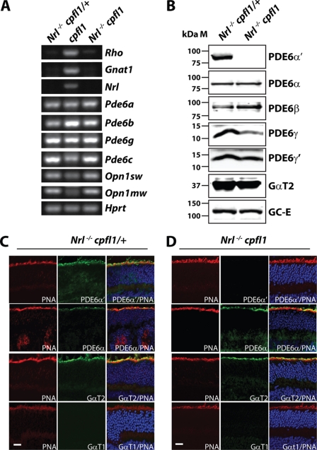
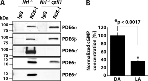
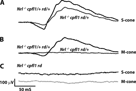
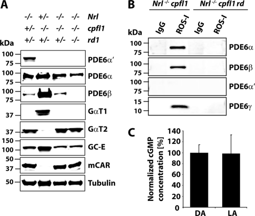
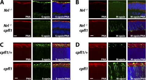
Rod and cone photoreceptor neurons utilize discrete PDE6 enzymes that are crucial for phototransduction. Rod PDE6 is composed of heterodimeric catalytic subunits (αβ), while the catalytic core of cone PDE6 (α') is a homodimer. It is not known if variations between PDE6 subunits preclude rod PDE6 catalytic subunits from coupling to the cone phototransduction pathway. To study this issue, we generated a cone-dominated mouse model lacking cone PDE6 (Nrl(-/-) cpfl1). In this animal model, using several independent experimental approaches, we demonstrated the expression of rod PDE6 (αβ) and the absence of cone PDE6 (α') catalytic subunits. The rod PDE6 enzyme expressed in cone cells is active and contributes to the hydrolysis of cGMP in response to light. In addition, rod PDE6 expressed in cone cells couples to the light signaling pathway to produce S-cone responses. However, S-cone responses and light-dependent cGMP hydrolysis were eliminated when the β-subunit of rod PDE6 was removed (Nrl(-/-) cpfl1 rd). We conclude that either rod or cone PDE6 can effectively couple to the cone phototransduction pathway to mediate visual signaling. Interestingly, we also found that functional PDE6 is required for trafficking of M-opsin to cone outer segments.


Figures

FIGURE 1.
FIGURE 1.
Light-dependent ERG. A, scotopic ERG measuring rod function in Nrl−/− cpfl1/+ and Nrl−/− cpfl1 mice at P30 (n = 3). As controls, we measured responses from C57Bl/6 and cpfl1 mice. The light intensity used to measure scotopic ERG was −0.8 log cd s/m2. B, photopic ERG measuring cone function in Nrl−/− cpfl1/+ and Nrl−/− cpfl1 mice at P30 (n = 3). C57bl/6 and cpfl1 mice serve as positive and negative controls, respectively. A typical response from each animal is shown. Photopic ERGs were measured at 0.4 log cd s/m2 xenon white flash with steady background light.
FIGURE 2.
FIGURE 2.
Cone-isolated ERGs. Light adapted ERGs from Nrl−/− cpfl1/+ (A) and Nrl−/− cpfl1 mice (B) at P30 to increasing light intensities of short wavelength monochromatic stimuli (360 nm) ranging from −3.6 to 0.7 log cd s/m2. Selected traces at the indicated light intensities are shown. Responses obtained from ERG recording were plotted against light intensities (C and D). Curves were fitted using Michaelis-Menten function as described under “Experimental Procedures.” Photoreceptor response (a-wave) and downstream bipolar response (b-wave) are depicted in panels C and D, respectively. Responses depicted are an average ± S.E. response from both eyes of three mice. M-cone ERG in Nrl−/− cpfl1/+ and Nrl−/− cpfl1 mice measured in response to monochromatic stimuli at 530 nm (E). Responses depicted are an average ± S.E. response from both eyes of three mice.
FIGURE 3.
FIGURE 3.
Expression of rod PDE6 in cone photoreceptor cells. RT-PCR analysis using retinal RNA extracted from in Nrl−/− cpfl1/+ and Nrl−/− cpfl1 mice at P12 (A). Expression of rod specific genes in the middle panel from retinal tissue lacking cone PDE6 (cpfl1) serves as positive control. Hprt, a housekeeping gene serves as loading control (A). Immunoblot analysis with indicated antibodies investigating the expression levels of proteins in retinal extracts from P30 Nrl−/− cpfl1/+ and Nrl−/− cpfl1 mice. Equal amounts (150 μg) of total proteins were loaded in each lane (B). Immunolocalization of rod and cone specific PDE6 and transducin in frozen retinal sections (P30) from Nrl−/− cpfl1/+ (C) and Nrl−/− cpfl1 mice (D). TO-PRO-3 stained nuclei are shown in blue, and peanut agglutinin (PNA)-stained cones are depicted in red. Cone PDE6α′, rod PDE6α, cone α-transducin (GαT2), and rod α-transducin (GαT1) staining are shown in green. (Scale bar: 10 μm.)
FIGURE 4.
FIGURE 4.
Rod PDE6 expressed in Nrl−/−cpfl1 mice is functionally active. A, immunoprecipitation (IP) of assembled rod PDE6 αβγ subunits from retinal extracts of Nrl−/− and Nrl−/− cpfl1 mice at P30 using ROS-I monoclonal antibody. After ROS-I IP, immunoblots were probed with rod or cone-specific PDE6 antibodies as indicated. Control IP with nonspecific mouse IgG is shown. B, amount of total cGMP, measured in dark (DA) and light-adapted (LA) retina from Nrl−/− cpfl1 mice. The data are presented as mean ± S.E. n = 3, *, p < 0.0017 compared with dark-adapted mice. Light-adapted retinas were obtained after mice (P30) were exposed to constant white light (71 cd/m2) in the ERG Ganzfeld for 15 min. Dark-adapted retinas were obtained from mice after overnight adaptation.
FIGURE 5.
FIGURE 5.
S-cone ERG is eliminated in Nrl−/− mice with defective rod and cone PDE6. Light-adapted S-cone responses in controls, Nrl−/− cpfl1/+ rd/+ (A), Nrl−/− cpfl1 rd/+ (A) and in mice lacking rod PDE6β subunit, Nrl−/− cpfl1 rd mice (C). M-cone responses in Nrl−/− cpfl1 rd/+ (B) and Nrl−/− cpfl1 rd (C) and in control, Nrl−/− cpfl1/+ rd/+ mice (B). All recordings were performed using littermate controls at P30 with light intensity of 0.7 log cd s/m2.
FIGURE 6.
FIGURE 6.
Rod PDE6 functions as an obligate heteromer. A, equal amounts (150 μg) of retinal extracts from littermate controls at P30 were used for immunoblot analysis with indicated antibodies. B, assessing the assembly of PDE6 by ROS-I. IP using mouse IgGs served as controls. Retina from P30 mice were used for these experiments. C, cGMP levels measured in the dark- (DA) and light-adapted (LA) retinas from Nrl−/− cpfl1 rd mice at P30. Light- and dark-adapted retinas were obtained as described earlier.
FIGURE 7.
FIGURE 7.
Functional PDE6 is crucial for localization of M-opsin to outer segments. Frozen retinal sections were probed with anti S-opsin (green) and peanut agglutinin (red). S-opsin is present in outer segments irrespective of the functional status of PDE6 (A, C). M -opsin (green) is present in outer segments in Nrl−/− and cpfl1/+ mice (B, D: upper panel) but is mislocalized to synaptic and nuclear layer of retina from Nrl−/− cpfl1 and cpfl1 mice (B, D: lower panel). All retinal sections were from P12 mice. (Scale bar: 10 μm.)

Similar articles

Cited by

References

    1. Pugh E. N., Jr., Cobbs W. H. (1986) Vision Res. 26, 1613–1643 - PubMed
    1. Fu Y., Yau K. W. (2007) Pflugers Arch 454, 805–819 - PMC - PubMed
    1. Burns M. E., Arshavsky V. Y. (2005) Neuron 48, 387–401 - PubMed
    1. Zhang X., Cote R. H. (2005) Front Biosci. 10, 1191–1204 - PubMed
    1. Kefalov V., Fu Y., Marsh-Armstrong N., Yau K. W. (2003) Nature 425, 526–531 - PMC - PubMed

Publication types

MeSH terms