Review
. 2011 Sep 23;286(38):32883-9.
doi: 10.1074/jbc.R110.197046. Epub 2011 Jul 28.

Allergic asthma: influence of genetic and environmental factors

Affiliations
Review

Allergic asthma: influence of genetic and environmental factors

Anil B Mukherjee et al. J Biol Chem. .

Abstract

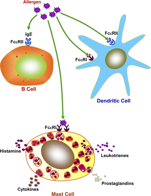
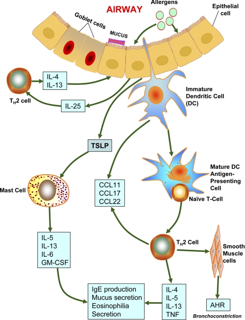
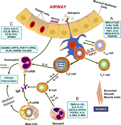
Allergic asthma is a chronic airway inflammatory disease in which exposure to allergens causes intermittent attacks of breathlessness, airway hyper-reactivity, wheezing, and coughing. Allergic asthma has been called a "syndrome" resulting from a complex interplay between genetic and environmental factors. Worldwide, >300 million individuals are affected by this disease, and in the United States alone, it is estimated that >35 million people, mostly children, suffer from asthma. Although animal models, linkage analyses, and genome-wide association studies have identified numerous candidate genes, a solid definition of allergic asthma has not yet emerged; however, such studies have contributed to our understanding of the multiple pathways to this syndrome. In contrast with animal models, in which T-helper 2 (T(H)2) cell response is the dominant feature, in human asthma, an initial exposure to allergen results in T(H)2 cell-dependent stimulation of the immune response that mediates the production of IgE and cytokines. Re-exposure to allergen then activates mast cells, which release mediators such as histamines and leukotrienes that recruit other cells, including T(H)2 cells, which mediate the inflammatory response in the lungs. In this minireview, we discuss the current understanding of how associated genetic and environmental factors increase the complexity of allergic asthma and the challenges allergic asthma poses for the development of novel approaches to effective treatment and prevention.


Figures

FIGURE 1.
FIGURE 1.
IgE-mediated allergic response. A wide variety of immune cells such as DCs and B cells express the low-affinity IgE receptor, FcϵRII (CD23). Uptake of allergen is mediated via IgE-bound FcϵRI and FcϵRII on APCs, augmenting secondary immune responses. The mast cells and basophils express the high-affinity IgE receptor, FcϵRI, which binds IgE, and the cross-linking of IgE-bound FcϵRI on these cells mediates release of proinflammatory mediators such as histamine, prostaglandins, leukotrienes, cytokines, and enzymes that lead to biological manifestation of allergy (8).
FIGURE 2.
FIGURE 2.
Possible pathways to allergic asthma. Allergens reaching the airways via inhaled air are taken up and processed by DCs that are primed by thymic stromal lymphopoietin (TSLP) secreted by airway epithelial cells. These allergens also cause the mast cells to release CCL17 and CCL22. CCL17 and CCL22 act on CCR4 (CC chemokine receptor 4), which mediates chemotactic migration of TH2 cells. TH2 cells play critical roles in orchestrating the allergen-induced inflammatory response by releasing IL-4 and IL-13. These interleukins also stimulate IgE production by B cells. These activated B cells also produce IL-5 (required for eosinophilic inflammation) and IL-9 (stimulator of mast cell proliferation). Airway epithelial cells release CCL11, stimulating recruitment of eosinophils via CCR3. Individuals suffering from allergic asthma may have defective Treg cells, which favor further TH2 cell proliferation and differentiation. Allergens also stimulate activation of sensitized mast cells by cross-linking surface-bound IgE molecules. In turn, activated mast cells secrete mediators of bronchoconstriction such as histamines, prostaglandin D2, and cysteinyl leukotrienes (2).
FIGURE 3.
FIGURE 3.
Genes identified by association studies or positional cloning. The genes that are associated with asthma/atopy are divided into four groups (26). A, the first group of genes is associated with triggering the allergic response via differentiation of CD4+ T helper cells. This group includes genes encoding CD14, TLR2, TLR4, TLR6, TLR10, NOD1, and NOD2, which are known as pattern recognition receptors. This group also includes genes that encode immunoregulatory cytokines such as IL-10 and TGF-β1; the transcription factor STAT3; antigen-presenting facilitator genes such as the HLA-DR, HLA-DQ, and HLA-DP alleles; and prostaglandin E receptor 2. B, the second group of genes includes GATA3, TBX21, IL4, IL13, IL4RA, FCER1B, IL5, IL5RA, STAT6, and IL12B, which regulate TH2 cell differentiation and effector functions. C, the third group includes genes encoding chemokines CCL5, CCL11, CCL24, and CCL26; antimicrobial peptide DEFB1; anti-inflammatory protein CC16 (also called UG); and factors responsible for maintaining the epithelial cell barrier such as SPINK5 and FLG. The positional cloning method has been used to identify the following genes expressed in the epithelia and smooth muscles: ADAM33, COL29A1, DPP10, GPRA, HLA-G, IRAKM, and PHF11 (26).

References

    1. Garantziotis S., Schwartz D. A. (2010) Annu. Rev. Public Health 31, 37–51 - PMC - PubMed
    1. Barnes P. J. (2008) Nat. Rev. Immunol. 8, 183–192 - PubMed
    1. Locksley R. M. (2010) Cell 140, 777–783 - PMC - PubMed
    1. Mannino D. M., Buist A. S. (2007) Lancet 370, 765–773 - PubMed
    1. Pearce N., Aït-Khaled N., Beasley R., Mallol J., Keil U., Mitchell E., Robertson C., and the ISAAC Phase Three Study Group (2007) Thorax 62, 758–766 - PMC - PubMed

Publication types