

Biometry of the corpus callosum in children: MR imaging reference data
- PMID: 21799035
- PMCID: PMC7964359
- DOI: 10.3174/ajnr.A2542
Biometry of the corpus callosum in children: MR imaging reference data
Abstract
Background and purpose: The availability of data relating to the biometry of the CC in children that are easy to use in daily practice is limited. We present a reference biometry of the CC in MR imaging in a large cohort of children.
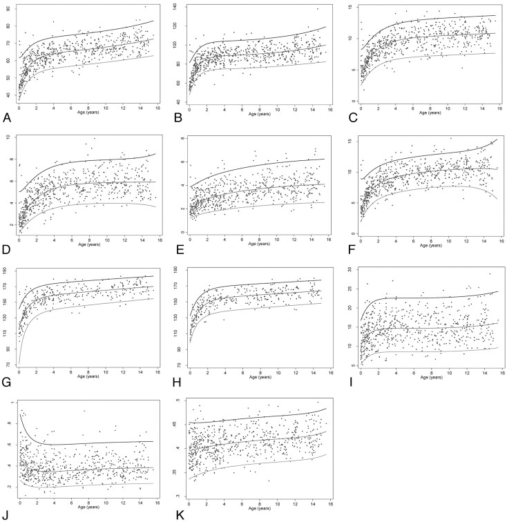
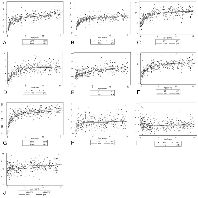
Materials and methods: Cerebral MR imaging studies of children with normal examination findings were selected retrospectively. Children born preterm and those with or at risk of developing cerebral malformations were excluded. The following parameters were measured: FOD, APD, LCC, GT, BT, IT, ST, and the S/T. Inter- and intraobserver agreement and sex effect were evaluated.
Results: Six hundred twenty-two children were included (320 boys, 302 girls), ranging from 1 day to 15 years of age. Normal values (from the 3rd to 97th percentile) are provided for each parameter. All parameters showed rapid growth up to 3 years of age followed by slower (FOD, APD, LCC, GT and ST) or absent (S/T) growth. Growth of BT and IT was completed by 7-8 years. CC modeling (IT/ST) was completed by 3 years. FOD was larger in boys from the age of 1 year (statistically significant). The other parameters did not show any sex effect. Inter- and intraobserver agreement was excellent for all parameters except for IT.
Conclusions: As measured, our data result in easy and reproducible MR imaging biometry of the CC in children.
Figures

References
-
- Royston P, Wright EM. A method for estimating age-specific reference intervals (‘normal ranges') based on fractional polynomials and exponential transformation. J R Statist Soc A 1998;161:79–101
-
- Royston P, Wright EM. Goodness-of-fit statistics for age-specific reference intervals. Stat Med 2000;19:2943–62 - PubMed
-
- Borghi E, de Onis M, Garza C, et al. . Construction of the World Health Organization child growth standards: selection of methods for attained growth curves. Stat Med 2006;25:247–65 - PubMed
-
- Rakic P, Yakovlev PI. Development of the corpus callosum and cavum septi in man. J Comp Neurol 1968;132:45–72 - PubMed
-
- Griffiths PD, Batty R, Reeves MJ, et al. . Imaging the corpus callosum, septum pellucidum and fornix in children: normal anatomy and variations of normality. Neuroradiology 2009;51:337–45 - PubMed
MeSH terms
LinkOut - more resources
Full Text Sources
Medical
Research Materials
Miscellaneous