. 2011 Nov-Dec;32(10):1926-9.
doi: 10.3174/ajnr.A2631. Epub 2011 Jul 28.

CT imaging features of obturator prostheses in patients following palatectomy or maxillectomy

Affiliations

CT imaging features of obturator prostheses in patients following palatectomy or maxillectomy

V A Kumar et al. AJNR Am J Neuroradiol. 2011 Nov-Dec.

Abstract

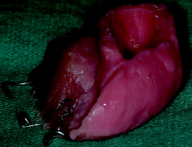
Palatal tumors are often treated with palatectomy or maxillectomy. The resulting surgical defect produces an oroantral communication. An obturator is a removable prosthesis used to close the palatal or maxillectomy defect. Fifteen patients who had undergone palatectomy or maxillectomy for carcinoma and subsequent obturator prosthesis placement were retrospectively studied. Obturators were characterized by Hounsfield units and were subdivided into 3 CT imaging groups: either hyperattenuated, hollow (air-containing), or heterogeneous (isoattenuated to hyperattenuated with internal foci of air). Eight patients had hyperattenuated obturators either representing acrylic resin or Trusoft. Four patients had hollow obturators also composed of acrylic resin or Trusoft. Three patients had heterogeneous obturators, which were composed of only Trusoft. The postoperative imaging of patients treated for palatal or maxillary tumors can be complicated by the presence of obturator prostheses. The intent of this article was to familiarize the reader with the CT imaging features of obturator prostheses.


Figures

Fig 1.
Fig 1.
A and B, A 32-year-old woman status post partial palatectomy for mucoepidermoid Ca and placement of a Trusoft obturator at the palatal defect (black arrow). The obturator bulb (white arrow) is seen extending to the level of the left choana. The bulb is the part of the obturator that inserts into the palatal defect. The postcontrast CT image shows the rounded hyperattenuated appearance of this obturator bulb, which was mistaken for enhancing tumor recurrence.
Fig 2.
Fig 2.
The interim obturator is constructed primarily of Trusoft and is formulated to remain soft for 6–10 weeks. The elasticity of Trusoft ensures against pressure pain. It is adjusted and contoured approximately every 2 weeks as the surgical site heals.
Fig 3.
Fig 3.
An 83-year-old woman with sinonasal lymphoma status post chemoradiation therapy, who developed osteoradionecrosis and underwent partial palatectomy and Trusoft obturator placement. A postcontrast CT image shows that the obturator projects into the right nasal cavity (arrow). Foci of air may get trapped in the Trusoft obturator, creating a confusing imaging appearance, which can be mistaken for infection.
Fig 4.
Fig 4.
Axial postcontrast CT images demonstrate 2 other examples of patients status post maxillectomy for tumor resection, with Trusoft obturators (arrows) containing trapped internal foci of air.
Fig 5.
Fig 5.
An example of a definitive obturator made of acrylic resin, with a closed hollow obturator bulb (arrow) molded to fill the palatal defect.
Fig 6.
Fig 6.
A 21-year-old woman with maxillary hemangioendothelioma status post left subtotal maxillectomy (left image) and subsequent acrylic obturator placement (right image with arrow), as shown on axial postcontrast CT images.
Fig 7.
Fig 7.
A 69-year-old patient with SCC of the right maxilla status post subtotal maxillectomy. An axial postcontrast CT image shows a hollow obturator (arrow) in place.

References

    1. Ortegon SM, Martin JW, Lewin JS. A hollow delayed surgical obturator for a bilateral subtotal maxillectomy patient: a clinical report. J Prosthet Dent 2008;99:14–18 - PubMed
    1. Keyf F. Obturator prostheses for hemimaxillectomy patients. J Oral Rehabil 2001;28:821–29 - PubMed
    1. Beumer J, III, Curtis TA, Marunick MT. Maxillofacial Rehabilitation: Prosthodontic and Surgical Considerations. St. Louis: Ishiyaku EuroAmerica; 1996:240–85