. 2011 Aug 23;108(34):14079-84.
doi: 10.1073/pnas.1108777108. Epub 2011 Jul 28.

Designed oligomers of cyanovirin-N show enhanced HIV neutralization

Affiliations

Designed oligomers of cyanovirin-N show enhanced HIV neutralization

Jennifer R Keeffe et al. Proc Natl Acad Sci U S A. .

Abstract

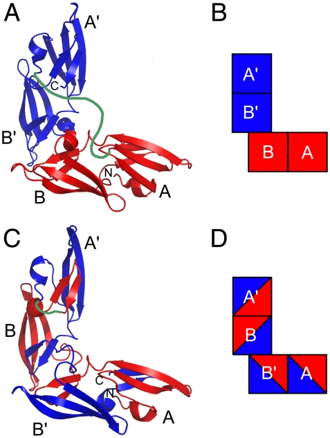
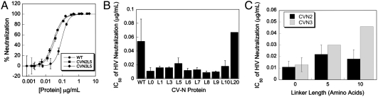
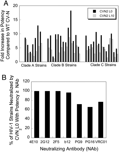
Cyanovirin-N (CV-N) is a small, cyanobacterial lectin that neutralizes many enveloped viruses, including human immunodeficiency virus type I (HIV-1). This antiviral activity is attributed to two homologous carbohydrate binding sites that specifically bind high mannose glycosylation present on envelope glycoproteins such as HIV-1 gp120. We created obligate CV-N oligomers to determine whether increasing the number of binding sites has an effect on viral neutralization. A tandem repeat of two CV-N molecules (CVN(2)) increased HIV-1 neutralization activity by up to 18-fold compared to wild-type CV-N. In addition, the CVN(2) variants showed extensive cross-clade reactivity and were often more potent than broadly neutralizing anti-HIV antibodies. The improvement in activity and broad cross-strain HIV neutralization exhibited by these molecules holds promise for the future therapeutic utility of these and other engineered CV-N variants.


Conflict of interest statement

The authors declare no conflict of interest.

Figures

Fig. 1.
Fig. 1.
Two models of CVN2 proteins. (A, B) The linked monomer dimeric model for CVN2s. (C, D) The linked domain-swapped dimeric model for CVN2s. The CV-N repeats are shown in red and blue, and the flexible polypeptide linker is modeled in green. The N and C termini are labeled N and C. Each of the four carbohydrate binding domains are labeled A, B, A′, or B′ in each model. Models (A) and (C) are based on solved WT CV-N structures (10, 44) and (B) and (D) are block representations of the models in (A) and (C), respectively.
Fig. 2.
Fig. 2.
CV-N oligomers exhibit enhanced HIV neutralization. (A) Typical neutralization data and curve fits for WT CV-N and two variants run in triplicate on the same 96-well plate. (B) The IC50s of HIV neutralization for WT CV-N and CVN2 dimers containing varying linker lengths. All linked dimers show significant enhancements in their HIV neutralization compared to WT CV-N, and dimers containing 0 to 10 amino acid linkers are more potent than CVN2L20. N≥4; CVN2L20: N = 1. (C) IC50s of HIV neutralization for CVN2s and CVN3s of the same linker length. N≥3; CVN3L5 and CVN3L10: N = 1. (A–C) Error bars = SD.
Fig. 3.
Fig. 3.
CV-N dimers neutralize HIV broadly and potency is similar to broadly neutralizing anti-HIV antibodies. (A) The designed dimers show enhanced neutralization activity relative to WT CV-N across all 33 strains tested from 3 clades. CVN2L0 is more potent than CVN2L10 in 32 of 33 cases. (B) When the IC50s of CVN2L0 neutralization against a panel of HIV-1 strains were compared to the IC50s of seven broadly neutralizing antibodies (Table S3), we saw that most strains were as sensitive to CVN2L0 as they were to the broadly neutralizing antibodies. Because 2-fold differences in potency are generally not significant, similar potency (≥) is defined as a potency for CVN2L0 that is within 2-fold of the potency of the antibody or higher.
Fig. 4.
Fig. 4.
Anti-HIV activity of CVN2L0 correlates with number of functional binding sites. (A) Schematic representation of variants to determine whether CVN2L0 is in a linked monomer dimeric structure (mm) or domain-swapped dimeric structure. The two CV-N repeats are represented in red and blue, as in Fig. 1 B and D. Black triangles represent partial carbohydrate binding site knockouts and black squares represent complete binding site knockouts. (B) HIV neutralization results for mutants in A. Mutants with full binding site deletions in the context of the domain-swapped dimer model have more significant increases in their HIV neutralization IC50s compared to mutants with full binding site deletions in the context of the monomer model. (C) Schematic representations of multiple binding site mutants. All variants contain one or more complete binding site knockouts according to the CVN2L0 domain-swapped dimer model. Black squares represent binding sites that have been knocked out, and squares containing red and blue triangles represent WT (functional) binding sites. (D) HIV neutralization results for mutants in C. The number of functional binding sites in CVN2L0 is proportional to its ability to neutralize HIV. Mutants with two functional binding sites are less active than those with three sites. Additionally, the deletion of a B binding site has a greater effect on activity than the deletion of an A binding site.
Fig. 5.
Fig. 5.
Similarity in carbohydrate binding site spacing in CV-N and the 2G12 anti-HIV (Fab)2. (A) Each of the four carbohydrate binding sites in one WT CV-N crystal structure (15) (P41212 space group) is approximately 30 to 35 Å from the other sites (structure is viewed from the bottom with respect to Fig. 1). Carbohydrates (shown as sticks with black carbons and red oxygens) were only resolved in the A binding sites in the crystal structure. (B) Ribbon diagram of the domain-swapped (Fab)2 from IgG 2G12, a broadly neutralizing antibody specific for carbohydrates on gp120 (35). The domain swapping creates a rigid (Fab)2 dimer in which the carbohydrate binding sites at the antigen combining sites are spaced approximately 30 to 35 Å apart. Carbohydrates are shown as sticks with black carbons and red oxygens and antibody domains are labeled.

References

    1. Boyd MR, et al. Discovery of cyanovirin-N, a novel human immunodeficiency virus-inactivating protein that binds viral surface envelope glycoprotein gp120: potential applications to microbicide development. Antimicrob Agents Chemother. 1997;41:1521–1530. - PMC - PubMed
    1. Dey B, et al. Multiple antiviral activities of cyanovirin-N: blocking of human immunodeficiency virus type 1 gp120 interaction with CD4 and coreceptor and inhibition of diverse enveloped viruses. J Virol. 2000;74:4562–4569. - PMC - PubMed
    1. O’Keefe BR, et al. Potent anti-influenza activity of cyanovirin-N and interactions with viral hemagglutinin. Antimicrob Agents Chemother. 2003;47:2518–2525. - PMC - PubMed
    1. Smee DF, et al. Influenza A (H1N1) virus resistance to cyanovirin-N arises naturally during adaptation to mice and by passage in cell culture in the presence of the inhibitor. Antivir Chem Chemoth. 2007;18:317–327. - PubMed
    1. Barrientos LG, Gronenborn AM. The highly specific carbohydrate-binding protein cyanovirin-N: structure, anti-HIV/Ebola activity and possibilities for therapy. Mini-Rev Med Chem. 2005;5:21–31. - PubMed

Publication types

MeSH terms

LinkOut - more resources