. 2011 Sep 16;109(7):783-93.
doi: 10.1161/CIRCRESAHA.111.243964. Epub 2011 Jul 28.

PGC-1β deficiency accelerates the transition to heart failure in pressure overload hypertrophy

Affiliations

PGC-1β deficiency accelerates the transition to heart failure in pressure overload hypertrophy

Christian Riehle et al. Circ Res. .

Abstract

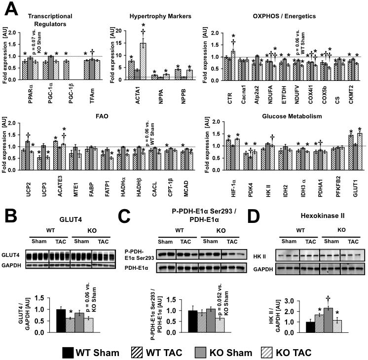
Rationale: Pressure overload cardiac hypertrophy, a risk factor for heart failure, is associated with reduced mitochondrial fatty acid oxidation (FAO) and oxidative phosphorylation (OXPHOS) proteins that correlate in rodents with reduced PGC-1α expression.

Objective: To determine the role of PGC-1β in maintaining mitochondrial energy metabolism and contractile function in pressure overload hypertrophy.

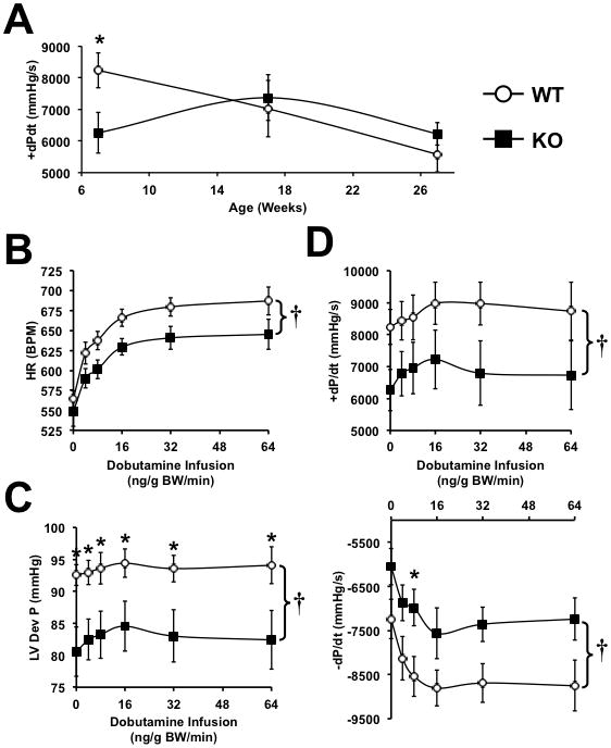
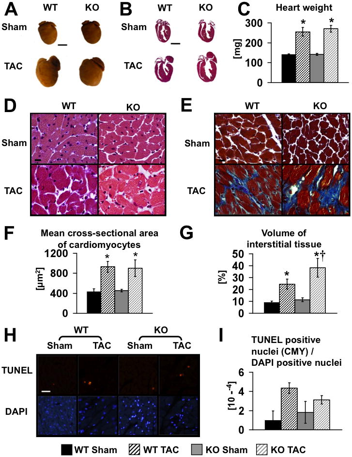
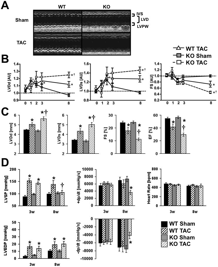
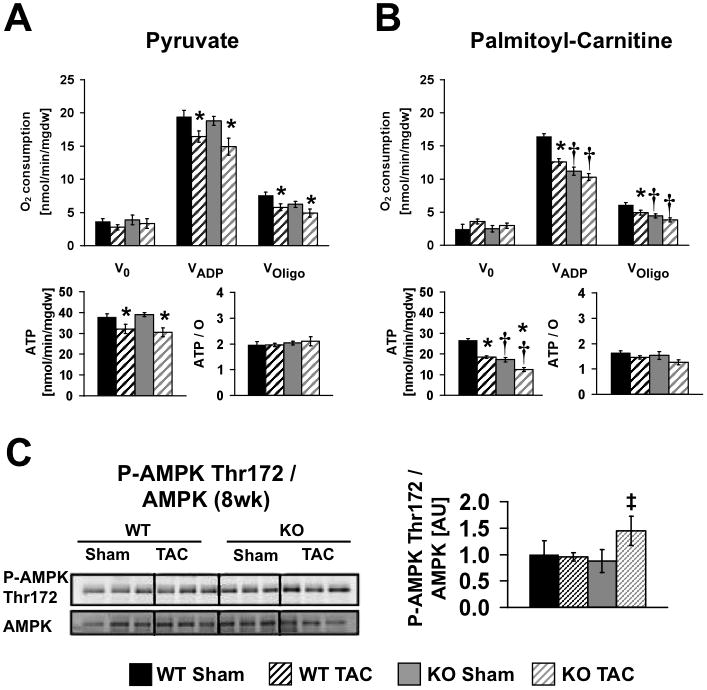
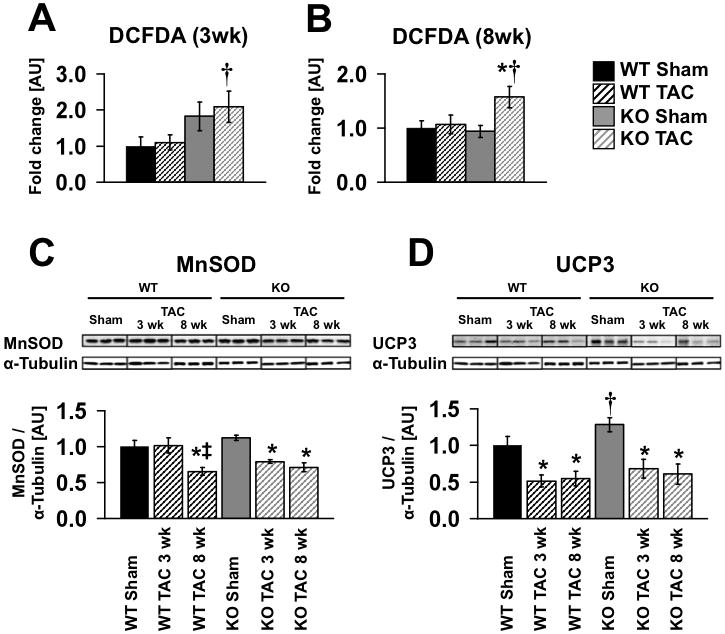
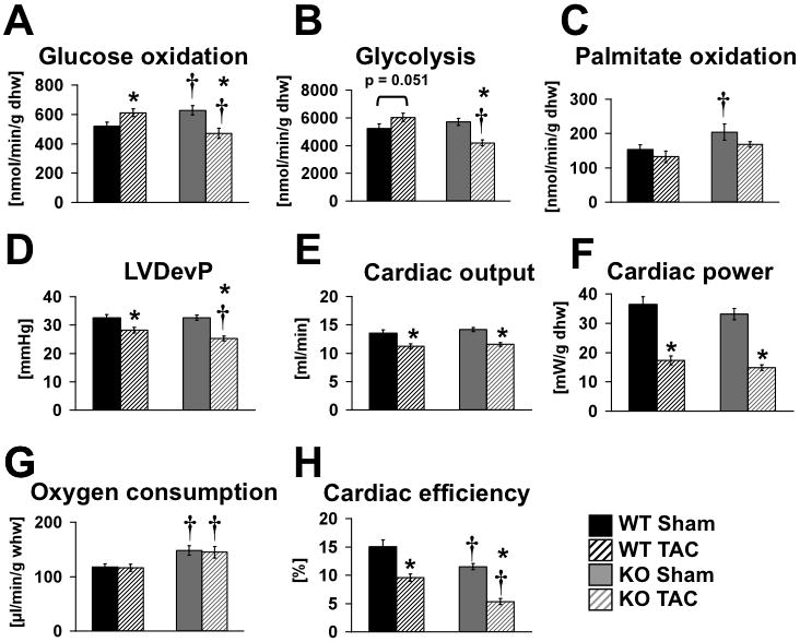
Methods and results: PGC-1β deficient (KO) mice and wildtype (WT) controls were subjected to transverse aortic constriction (TAC). Although LV function was modestly reduced in young KO hearts, there was no further decline with age so that LV function was similar between KO and WT when TAC was performed. WT-TAC mice developed relatively compensated LVH, despite reduced mitochondrial function and repression of OXPHOS and FAO genes. In nonstressed KO hearts, OXPHOS gene expression and palmitoyl-carnitine-supported mitochondrial function were reduced to the same extent as banded WT, but FAO gene expression was normal. Following TAC, KO mice progressed more rapidly to heart failure and developed more severe mitochondrial dysfunction, despite a similar overall pattern of repression of OXPHOS and FAO genes as WT-TAC. However, in relation to WT-TAC, PGC-1β deficient mice exhibited greater degrees of oxidative stress, decreased cardiac efficiency, lower rates of glucose metabolism, and repression of hexokinase II protein.

Conclusions: PGC-1β plays an important role in maintaining baseline mitochondrial function and cardiac contractile function following pressure overload hypertrophy by preserving glucose metabolism and preventing oxidative stress.


Conflict of interest statement

Disclosures

C. Lelliott is employed by Astra Zeneca. The other authors have no conflicts of interest to disclose.

Figures

Figure 1
Figure 1
(A) Age-dependent changes in peak rate of ventricular contraction (+dP/dt) in non-stressed WT and PGC-1β−/− hearts (n=5–9). (B–D) In vivo hemodynamic parameters in 7-week-old WT and PGC-1β−/− hearts following graded dobutamine infusion as indicated. * = p<0.05 vs. WT at equivalent dobutamine dose, † = p<0.05 for analysis of covariance comparing genotypes (n=5–7). Data were obtained in male and female mice and are combined.
Figure 2
Figure 2
(A) Representative photographs of male WT and KO hearts following eight weeks of TAC or Sham surgery (age 16–18 weeks). Scale bars, 3 mm. (B) Hematoxylin-eosin stains of longitudinal sections of the same hearts as in A. Scale bars, 3 mm. (C) Mean heart weights eight weeks after surgery, n=12. Hematoxylin-eosin (D) and trichrome stains (E) (scale bars, 10 μm) and quantification of mean cross-sectional area of cardiomyocytes (F) and volume of interstitial fibrosis (G). Representative TUNEL (upper) and DAPI (lower) staining (H) and quantification (I); Scale bar, 50 μm; CMY – cardiomyocytes. Data were obtained from 3 to 7 hearts per group. * = p<0.05 vs. Sham same genotype, † = p<0.05 vs. WT same treatment.
Figure 3
Figure 3
(A) Representative M-Mode echocardiographs from Sham (upper) or TAC (lower) male WT and KO mice illustrating cardiac contractile dysfunction in PGC-1β hearts after eight weeks of pressure overload (same mice as figure 2). IVS=interventricular septum, LVD=left ventricular diameter, LVPW=LV posterior wall; (B) Time course for LVDd - Left ventricular cavity diameter at diastole, LVDs - Left ventricular cavity diameter at systole, and FS - Fractional shortening presented as fold change vs. WT Sham (= 1.0). The X-axes show the time in weeks after surgery. (C) LVDd, LVDs, FS, and EF - Ejection fraction 8-weeks after Sham or TAC. (D) In vivo, left ventricular hemodynamic parameters, three and eight weeks after Sham or TAC surgery. * = p<0.05 vs. Sham same genotype, † = p<0.05 vs. WT same treatment (n = 6–13). Week 1–3 mice were banded at 23–30 weeks of age and week 8 data are from 16–18-week-old mice
Figure 4
Figure 4
Mitochondrial function of male and female WT and KO hearts (age 16–18 weeks) following eight weeks of TAC or Sham surgery (4 to 7 hearts per group). Mitochondrial respiration, ATP synthesis rates and ATP/O ratios were measured in saponin-permeabilized cardiac fibers. Pyruvate-malate (A) or palmitoyl-carnitine-malate (B) were used as substrates. (C) Western blot analysis and densitometric ratios of AMPK phosphorylation eight weeks post surgery (n=5–6). Data are presented as fold change vs. WT Sham (=1.0). * = p<0.05 vs. Sham same genotype, † = p<0.05 vs. WT same treatment ‡ = p<0.05 vs. KO Sham (Wilcoxon test)
Figure 5
Figure 5
Reactive oxygen species levels were measured by 2′-7′-dichlorofluorescein diacetate (DCFDA) fluorescence in whole tissue extract three (A) and eight (B) weeks after Sham or TAC surgery, n=4–7. (C, D) Representative western blots showing protein levels of MnSOD and UCP3 three and eight weeks after surgery and densitometric analysis normalized to α-tubulin (n = 5–11). Data are presented as fold change vs. WT Sham. * = p<0.05 vs. Sham same genotype, † = p<0.05 vs. WT same treatment, ‡ = p<0.05 vs. same genotype three weeks post surgery. TAC 3wk and TAC 8 wk were 8 and 13-weeks of age respectively at the time of sacrifice.
Figure 6
Figure 6
(A–H) Cardiac substrate metabolism and function in isolated working hearts. Glucose oxidation, glycolysis, palmitate oxidation, aortic developed pressure (DevP), cardiac output, cardiac power, oxygen consumption and cardiac efficiency in isolated working hearts isolated from wildtype (WT) and PGC-1β KO (KO) mice three weeks after TAC or Sham surgery, performed in mice between the ages of 23–30 weeks. Hearts were perfused with 5mM glucose and 0.4mM palmitate. n=4 hearts per group for metabolism, MVO2 and efficiency and 8/group for cardiac function (pooled data from glucose and FA perfusions). * = p<0.05 vs. Sham same genotype, † = p<0.05 vs. WT same treatment.
Figure 7
Figure 7
(A) Gene expression eight weeks after surgery (n = 8, same mice as Figure 2). (B, C, D) Western blot analysis and densitometric ratios of GLUT4 protein expression, PDH-E1α phosphorylation and Hexokinase II protein expression in WT and KO hearts eight weeks post surgery (n = 5–6, same mice as Figure 5). * = p<0.05 vs. Sham same genotype, † = p<0.05 vs. WT same treatment. Gene names are shown in Online Table III.

References

    1. Meirhaeghe A, Crowley V, Lenaghan C, Lelliott C, Green K, Stewart A, Hart K, Schinner S, Sethi JK, Yeo G, Brand MD, Cortright RN, O’Rahilly S, Montague C, Vidal-Puig AJ. Characterization of the human, mouse and rat pgc1 beta (peroxisome-proliferator-activated receptor-gamma co-activator 1 beta) gene in vitro and in vivo. Biochem J. 2003;373:155–165. - PMC - PubMed
    1. Lin J, Tarr PT, Yang R, Rhee J, Puigserver P, Newgard CB, Spiegelman BM. Pgc-1beta in the regulation of hepatic glucose and energy metabolism. J Biol Chem. 2003;278:30843–30848. - PubMed
    1. Fan M, Rhee J, St-Pierre J, Handschin C, Puigserver P, Lin J, Jaeger S, Erdjument-Bromage H, Tempst P, Spiegelman BM. Suppression of mitochondrial respiration through recruitment of p160 myb binding protein to pgc-1alpha: Modulation by p38 mapk. Genes Dev. 2004;18:278–289. - PMC - PubMed
    1. Lehman JJ, Boudina S, Banke NH, Sambandam N, Han X, Young DM, Leone TC, Gross RW, Lewandowski ED, Abel ED, Kelly DP. The transcriptional coactivator pgc-1alpha is essential for maximal and efficient cardiac mitochondrial fatty acid oxidation and lipid homeostasis. Am J Physiol Heart Circ Physiol. 2008;295:H185–196. - PMC - PubMed
    1. Garnier A, Fortin D, Delomenie C, Momken I, Veksler V, Ventura-Clapier R. Depressed mitochondrial transcription factors and oxidative capacity in rat failing cardiac and skeletal muscles. J Physiol. 2003;551:491–501. - PMC - PubMed

Publication types

MeSH terms