

Autoimmune responses to the brain after stroke are associated with worse outcome
- PMID: 21799171
- PMCID: PMC3183403
- DOI: 10.1161/STROKEAHA.111.619593
Autoimmune responses to the brain after stroke are associated with worse outcome
Abstract
Background and purpose: Immune responses to brain antigens occur after stroke, and experimental studies show that the likelihood of developing a detrimental autoimmune response to these antigens is increased by systemic inflammation at the time of stroke. The aim of this study was to determine if patients who developed infection in the poststroke period would be similarly predisposed to develop autoimmune responses to central nervous system antigens.
Methods: We enrolled 114 patients within 72 hours of ischemic stroke. Clinical and demographic data were obtained, and cellular immune responses to a panel of central nervous system antigens were assessed during the initial week and again at Day 90. Outcome was assessed using the modified Rankin Scale.
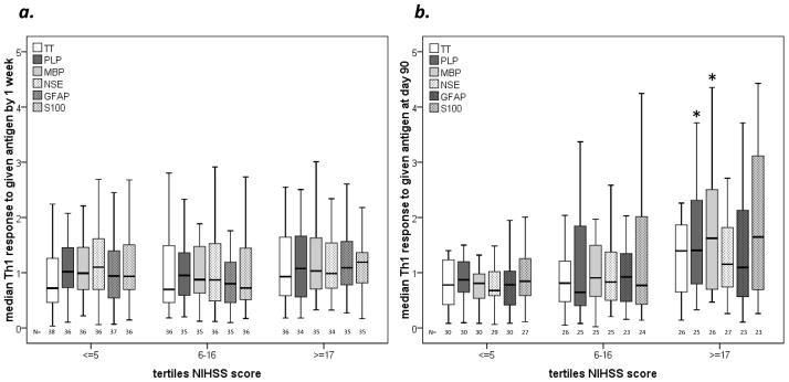
Results: Patients who developed an infection, especially pneumonia, in the 15 days after stroke were more likely to evidence a Th1(+) response to myelin basic protein and glial fibrillary acidic protein (P=0.019 and P=0.039, respectively) at 90 days after stroke. Further, more robust Th1 responses to myelin basic protein at 90 days were associated with a decreased likelihood of good outcome, even after adjusting for baseline stroke severity and patient age (OR, 0.477; 95% CI, 0.244 to 0.935; P=0.031).
Conclusions: This study demonstrates that immune responses to brain antigens occur after stroke. Although these responses are likely to be an epiphenomenon of ischemic brain injury, the response to myelin basic protein appears to have clinical consequences. The potential role of postischemic autoimmune-mediated brain injury deserves further investigation.
Figures
Comment in
-
Letter by Urra et al regarding article, "Autoimmune responses to the brain after stroke are associated with worse outcome".Stroke. 2012 Feb;43(2):e26; author reply e27-8. doi: 10.1161/STROKEAHA.111.643460. Epub 2012 Jan 5. Stroke. 2012. PMID: 22223242 No abstract available.
References
-
- Schroeter M, Jander S, Witte OW, Stoll G. Local immune responses in the rat cerebral cortex after middle cerebral artery occlusion. J Neuroimmunol. 1994;55:195–203. - PubMed
-
- Jander S, Kraemer M, Schroeter M, Witte OW, Stoll G. Lymphocytic infiltration and expression of intercellular adhesion molecule-1 in photochemically induced ischemia of the rat cortex. J Cereb Blood Flow Metab. 1995;15:42–51. - PubMed
-
- Braun JS, Jander S, Schroeter M, Witte OW, Stoll G. Spatiotemporal relationship of apoptotic cell death to lymphomonocytic infiltration in photochemically induced focal ischemia of the rat cerebral cortex. Acta Neuropathol (Berl) 1996;92:255–263. - PubMed
-
- Campanella M, Sciorati C, Tarozzo G, Beltramo M. Flow cytometric analysis of inflammatory cells in ischemic rat brain. Stroke. 2002;33:586–592. - PubMed
-
- Jauch EC, Lindsell C, Broderick J, Fagan SC, Tilley BC, Levine SR. Association of serial biochemical markers with acute ischemic stroke: The national institute of neurological disorders and stroke recombinant tissue plasminogen activator stroke study. Stroke. 2006;37:2508–2513. - PubMed
Publication types
MeSH terms
Grants and funding
LinkOut - more resources
Full Text Sources
Medical
