

Abnormal cortical thickness alterations in fetal alcohol spectrum disorders and their relationships with facial dysmorphology
- PMID: 21799209
- PMCID: PMC3328347
- DOI: 10.1093/cercor/bhr193
Abnormal cortical thickness alterations in fetal alcohol spectrum disorders and their relationships with facial dysmorphology
Abstract
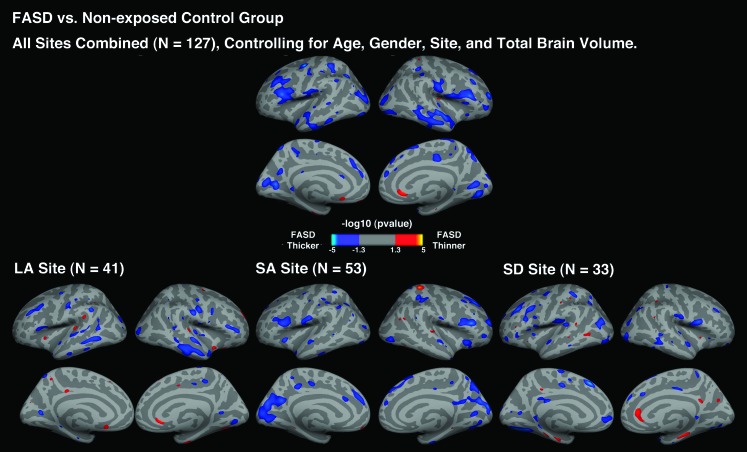
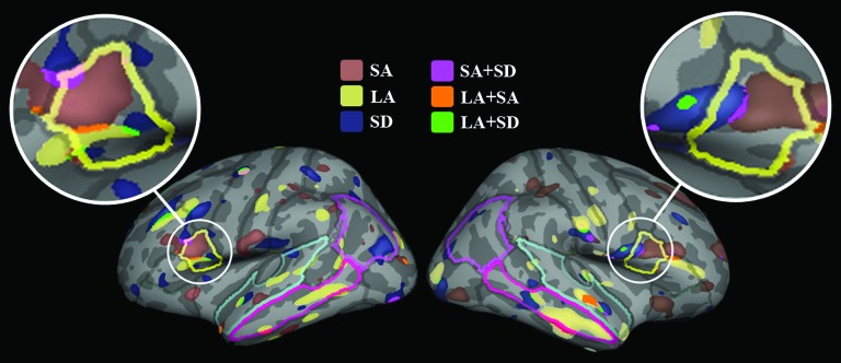
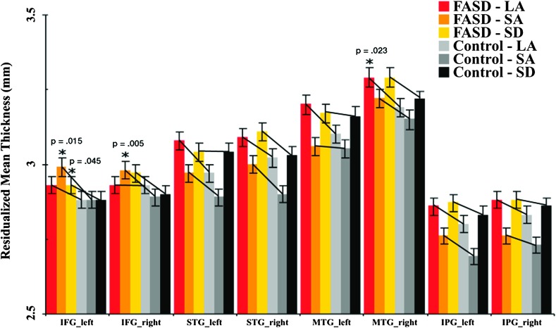
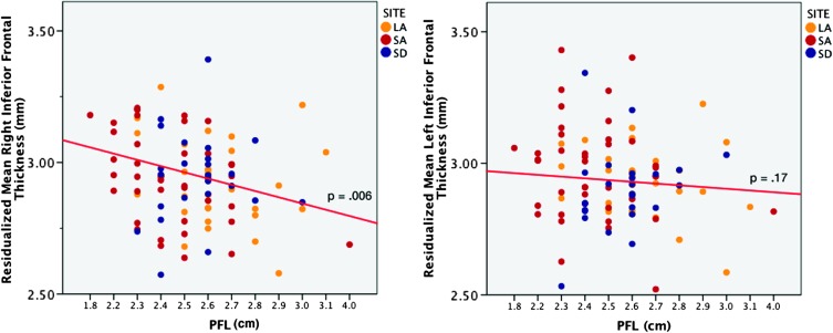
Accumulating evidence from structural brain imaging studies on individuals with fetal alcohol spectrum disorder (FASD) has supported links between prenatal alcohol exposure and brain morphological deficits. Although global and regional volumetric reductions appear relatively robust, the effects of alcohol exposure on cortical thickness and relationships with facial dysmorphology are not yet known. The structural magnetic resonance imaging data from 69 children and adolescents with FASD and 58 nonexposed controls collected from 3 sites were examined using FreeSurfer to detect cortical thickness changes across the entire brain in FASD and their associations with facial dysmorphology. Controlling for brain size, subjects with FASD showed significantly thicker cortices than controls in several frontal, temporal, and parietal regions. Analyses conducted within site further revealed prominent group differences in left inferior frontal cortex within all 3 sites. In addition, increased inferior frontal thickness was significantly correlated with reduced palpebral fissure length. Consistent with previous reports, findings of this study are supportive of regional increases in cortical thickness serving as a biomarker for disrupted brain development in FASD. Furthermore, the significant associations between thickness and dysmorphic measures suggest that the severity of brain anomalies may be reflected by that of the face.
Figures
Similar articles
-
Abnormal cortical thickness and brain-behavior correlation patterns in individuals with heavy prenatal alcohol exposure.Cereb Cortex. 2008 Jan;18(1):136-44. doi: 10.1093/cercor/bhm039. Epub 2007 Apr 18. Cereb Cortex. 2008. PMID: 17443018 Free PMC article.
-
Callosal thickness reductions relate to facial dysmorphology in fetal alcohol spectrum disorders.Alcohol Clin Exp Res. 2012 May;36(5):798-806. doi: 10.1111/j.1530-0277.2011.01679.x. Epub 2011 Dec 7. Alcohol Clin Exp Res. 2012. PMID: 22150665 Free PMC article.
-
Microstructural corpus callosum anomalies in children with prenatal alcohol exposure: an extension of previous diffusion tensor imaging findings.Alcohol Clin Exp Res. 2009 Oct;33(10):1825-35. doi: 10.1111/j.1530-0277.2009.01021.x. Epub 2009 Jul 23. Alcohol Clin Exp Res. 2009. PMID: 19645729 Free PMC article.
-
Neuroimaging and fetal alcohol spectrum disorders.Dev Disabil Res Rev. 2009;15(3):209-17. doi: 10.1002/ddrr.72. Dev Disabil Res Rev. 2009. PMID: 19731391 Free PMC article. Review.
-
Prenatal alcohol exposure and associations with physical size, dysmorphology and neurodevelopment: a systematic review and meta-analysis.BMC Med. 2024 Oct 15;22(1):467. doi: 10.1186/s12916-024-03656-w. BMC Med. 2024. PMID: 39407296 Free PMC article.
Cited by
-
Executive function deficits and social-behavioral abnormality in mice exposed to a low dose of dioxin in utero and via lactation.PLoS One. 2012;7(12):e50741. doi: 10.1371/journal.pone.0050741. Epub 2012 Dec 12. PLoS One. 2012. PMID: 23251380 Free PMC article.
-
Maternal choline supplementation in a sheep model of first trimester binge alcohol fails to protect against brain volume reductions in peripubertal lambs.Alcohol. 2016 Sep;55:1-8. doi: 10.1016/j.alcohol.2016.07.004. Epub 2016 Aug 4. Alcohol. 2016. PMID: 27788773 Free PMC article.
-
Cortical miscommunication after prenatal exposure to alcohol.Exp Brain Res. 2016 Nov;234(11):3347-3353. doi: 10.1007/s00221-016-4732-3. Epub 2016 Aug 4. Exp Brain Res. 2016. PMID: 27491551
-
Volume changes and brain-behavior relationships in white matter and subcortical gray matter in children with prenatal alcohol exposure.Hum Brain Mapp. 2015 Jun;36(6):2318-29. doi: 10.1002/hbm.22772. Epub 2015 Feb 25. Hum Brain Mapp. 2015. PMID: 25711175 Free PMC article.
-
Diffusion MRI of the developing cerebral cortical gray matter can be used to detect abnormalities in tissue microstructure associated with fetal ethanol exposure.Neuroimage. 2013 Dec;83:1081-7. doi: 10.1016/j.neuroimage.2013.07.068. Epub 2013 Aug 3. Neuroimage. 2013. PMID: 23921100 Free PMC article.
References
-
- Archibald SL, Fennema-Notestine C, Gamst A, Riley EP, Mattson SN, Jernigan TL. Brain dysmorphology in individuals with severe prenatal alcohol exposure. Dev Med Child Neurol. 2001;43:148–154. - PubMed
-
- Aron AR, Fletcher PC, Bullmore ET, Sahakian BJ, Robbins TW. Stop-signal inhibition disrupted by damage to right inferior frontal gyrus in humans. Nat Neurosci. 2003;6:115–116. - PubMed
-
- Aron AR, Robbins TW, Poldrack RA. Inhibition and the right inferior frontal cortex. Trends Cogn Sci. 2004;8:170–177. - PubMed
-
- Astley SJ, Aylward EH, Olsen HC, Kerns K, Brooks A, Coggins TE, Davies J, Dorn S, Gendler B, Jirikowic T, et al. Magnetic resonance imaging outcomes from a comprehensive magnetic resonance study of children with fetal alcohol spectrum disorders. Alcohol Clin Exp Res. 2009;33:1671–1689. - PMC - PubMed
Publication types
MeSH terms
Grants and funding
LinkOut - more resources
Full Text Sources
Medical
