

Abnormal cortical thickness alterations in fetal alcohol spectrum disorders and their relationships with facial dysmorphology
- PMID: 21799209
- PMCID: PMC3328347
- DOI: 10.1093/cercor/bhr193
Abnormal cortical thickness alterations in fetal alcohol spectrum disorders and their relationships with facial dysmorphology
Abstract
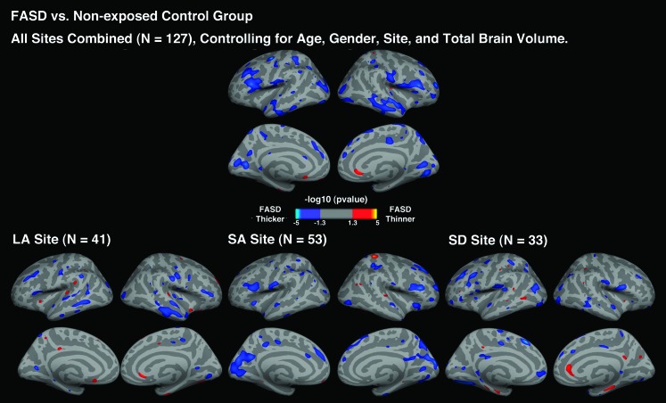
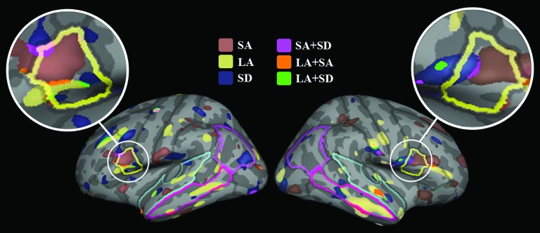
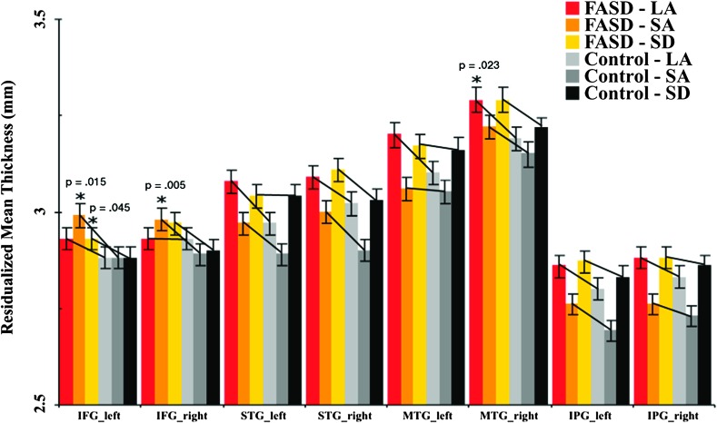
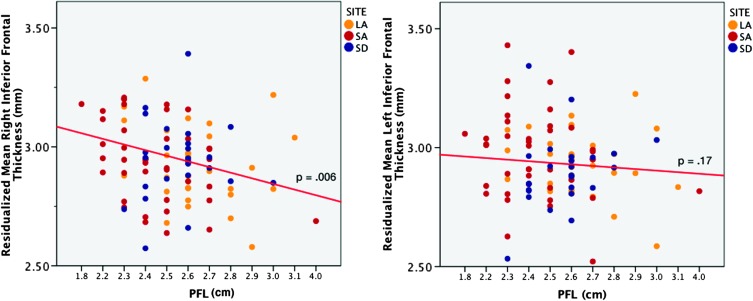
Accumulating evidence from structural brain imaging studies on individuals with fetal alcohol spectrum disorder (FASD) has supported links between prenatal alcohol exposure and brain morphological deficits. Although global and regional volumetric reductions appear relatively robust, the effects of alcohol exposure on cortical thickness and relationships with facial dysmorphology are not yet known. The structural magnetic resonance imaging data from 69 children and adolescents with FASD and 58 nonexposed controls collected from 3 sites were examined using FreeSurfer to detect cortical thickness changes across the entire brain in FASD and their associations with facial dysmorphology. Controlling for brain size, subjects with FASD showed significantly thicker cortices than controls in several frontal, temporal, and parietal regions. Analyses conducted within site further revealed prominent group differences in left inferior frontal cortex within all 3 sites. In addition, increased inferior frontal thickness was significantly correlated with reduced palpebral fissure length. Consistent with previous reports, findings of this study are supportive of regional increases in cortical thickness serving as a biomarker for disrupted brain development in FASD. Furthermore, the significant associations between thickness and dysmorphic measures suggest that the severity of brain anomalies may be reflected by that of the face.
Figures
References
-
- Archibald SL, Fennema-Notestine C, Gamst A, Riley EP, Mattson SN, Jernigan TL. Brain dysmorphology in individuals with severe prenatal alcohol exposure. Dev Med Child Neurol. 2001;43:148–154. - PubMed
-
- Aron AR, Fletcher PC, Bullmore ET, Sahakian BJ, Robbins TW. Stop-signal inhibition disrupted by damage to right inferior frontal gyrus in humans. Nat Neurosci. 2003;6:115–116. - PubMed
-
- Aron AR, Robbins TW, Poldrack RA. Inhibition and the right inferior frontal cortex. Trends Cogn Sci. 2004;8:170–177. - PubMed
-
- Astley SJ, Aylward EH, Olsen HC, Kerns K, Brooks A, Coggins TE, Davies J, Dorn S, Gendler B, Jirikowic T, et al. Magnetic resonance imaging outcomes from a comprehensive magnetic resonance study of children with fetal alcohol spectrum disorders. Alcohol Clin Exp Res. 2009;33:1671–1689. - PMC - PubMed
Publication types
MeSH terms
Grants and funding
LinkOut - more resources
Full Text Sources
Medical
