. 2011;27(1):211-23.
doi: 10.3233/JAD-2011-110209.

Coenzyme Q10 decreases amyloid pathology and improves behavior in a transgenic mouse model of Alzheimer's disease

Affiliations

Coenzyme Q10 decreases amyloid pathology and improves behavior in a transgenic mouse model of Alzheimer's disease

Magali Dumont et al. J Alzheimers Dis. 2011.

Abstract

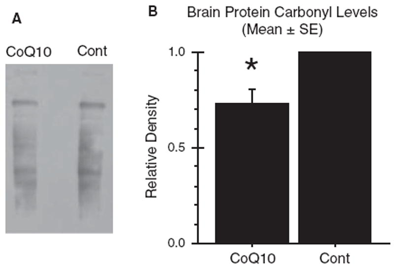
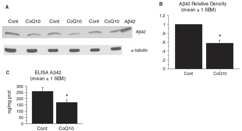
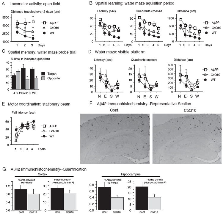
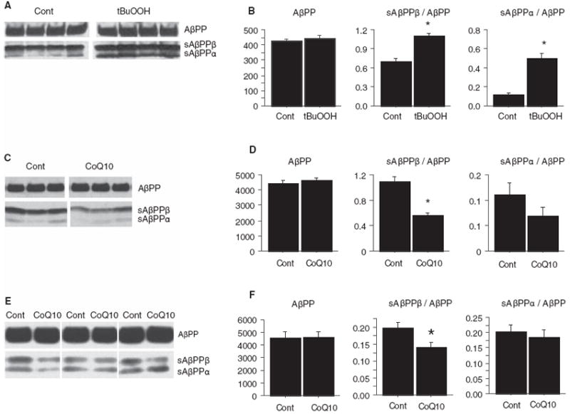
Increased oxidative stress is implicated in the pathogenesis of Alzheimer's disease (AD). A large body of evidence suggests that mitochondrial dysfunction and increased reactive oxygen species occur prior to amyloid-β (Aβ) deposition. Coenzyme Q10 (CoQ10), a component of the mitochondrial electron transport chain, is well characterized as a neuroprotective antioxidant in animal models and human trials of Huntington's disease and Parkinson's disease, and reduces plaque burden in AβPP/PS1 mice. We now show that CoQ10 reduces oxidative stress and amyloid pathology and improves behavioral performance in the Tg19959 mouse model of AD. CoQ10 treatment decreased brain levels of protein carbonyls, a marker of oxidative stress. CoQ10 treatment resulted in decreased plaque area and number in hippocampus and in overlying cortex immunostained with an Aβ42-specific antibody. Brain Aβ42 levels were also decreased by CoQ10 supplementation. Levels of amyloid-β protein precursor (AβPP) β-carboxyterminal fragments were decreased. Importantly, CoQ10-treated mice showed improved cognitive performance during Morris water maze testing. Our results show decreased pathology and improved behavior in transgenic AD mice treated with the naturally occurring antioxidant compound CoQ10. CoQ10 is well tolerated in humans and may be promising for therapeutic trials in AD.


Figures

Fig. 1
Fig. 1
Dietary supplementation with coenzyme Q10 reduced brain oxidative stress. Tg19959 mice were fed for three months with control chow or chow supplemented with 0.4% CoQ10 (800 mg/kg/day). A) Oxidative stress was assessed by Western blotting for protein carbonyls in 6% SDS brain homogenates. B) Densitometry. Samples were run in pairs, with one CoQ10-fed animal and one control-fed animal in each pair. In each pair, the total carbonyl densities from both the CoQ10-fed animal and the control-fed animal were divided by that of the control-fed animal. The graph shows the mean ± SEM for these ratios. *p = 0.0017, two-tailed t-test. n = 11 pairs were run.
Fig. 2
Fig. 2
Dietary supplementation with coenzyme Q10 decreased amyloid plaque area and number. Tg19959 mice were fed for three months with control chow or chow supplemented with 0.4% CoQ10 (800 mg/kg/day). A) Immunohistochemistry. Representative coronal section through retrosplenial cortex and hippocampus are shown. Sections were stained with polyclonal antibody AB5078P directed against Aβ42. B-E) Quantification of percent area occupied by plaque (B, D) and number of plaques per unit area (C, E) in cortex (B, C) and hippocampus (D, E). For each mouse, the plaque area and number were averaged over 5 sections, and the graphs show the means and standard errors of these averages over all mice. P-values were obtained by two-tailed unpaired t-test. For the quantification, n = 12 mice (6 male, 6 female) were used in each group. *p < 0.05.
Fig. 3
Fig. 3
Dietary supplementation with coenzyme Q10 decreased brain Aβ levels. Tg19959 mice were fed for three months with control chow or chow supplemented with 0.4% CoQ10 (800 mg/kg/day). A) Brain Aβ42 levels were assessed by western blotting the 6% SDS fraction of brain homogenates. Blots were probed with AB5078P (Chemicon, Temecula, CA). Forty μg protein was loaded in each well, and α-tubulin was used as a loading control. Right lane: Aβ42 standard. B) Densitometry. Samples were run in pairs, with one CoQ10-fed animal and one control-fed animal in each pair. In each pair, the density of the Aβ bands from both the CoQ10-fed animal and the control-fed animal were divided by that of the control-fed animal. The graph shows the mean ± SEM for these ratios. *p < 0.0001, two-tailed t-test. N= 17 pairs were run. C) Brain Aβ42 levels were also assessed by Sandwich ELISA using the 6% SDS fraction of brain homogenates. *p = 0.03, two-tailed t-test. N = 10 pairs were run.
Fig. 4
Fig. 4
Dietary supplementation with coenzyme Q10 improved behavioral performance. Tg19959 mice were fed for five months with control chow or chow supplemented with 2.4% CoQ10 (4800 mg/kg/day). For all measures in (A-E), number of mice used was: n = 6, CoQ10-fed Tg19959 (CoQ10); n = 12, control-fed Tg19959 (AβPP); n = 9, wild type littermate mice (WT). ANOVA with repetitive measurements were conducted for behavioral analyses. A) Open field test. Control-fed Tg19959 (AβPP) mice were hyperactive compared to wild-type (WT) mice when placed in the open field, as shown by the distance traveled each day (p = 0.01, AβPP versus WT). There was a trend for CoQ10-fed Tg19959 (CoQ10) mice to be similarly hyperactive compared to WT (CoQ10 versus WT, p = 0.30), but less so than control-fed Tg19959 mice (CoQ10 versus AβPP, p = 0.19). CoQ10-fed Tg19959 mice were able to habituate in the open field (note decrease in distance traveled on days 2 and 3 compared to day 1). In contrast, the control fed Tg19959 mice did not show any habituation. B) The Morris water maze was used to test spatial learning. Over the 5 day learning period with the hidden platform, control-fed Tg19959 mice (AβPP) performed worse than wild-type mice (WT) with respect to latency to reach platform, quadrants crossed, and distance traveled (p < 0.0001 for all measures). Performance of the CoQ10-fed Tg19959 mice was better than that of the control fed Tg19959 mice in all measures (latency, p = 0.0389; quadrants, p = 0.0014; distance, p = 0.0081), although not quite at the level of wild type mice (CoQ10 versus WT latency, p = 0.0227; quadrants, p = 0.1745; distance, p = 0.0978). C) Spatial memory was assessed 24 h after the acquisition period ended. The mice were released into the water maze with the platform removed (probe trial), and the percentage of time spent in the target quadrant was compared to that spent in the diagonally opposite quadrant. The control-fed mice Tg19959 mice showed no preference for the target quadrant; there was even a trend to prefer the opposite quadrant (p = 0.13). In contrast, the CoQ10-fed Tg19959 mice and the WT mice preferred the target over the opposite quadrant (p = 0.033 CoQ10; p = 0.0011 WT). (D) To ensure that CoQ10-supplementation did not confer a noncognitive advantage in swimming or vision, ability to reach a visible platform was measured. CoQ10fed Tg19959 mice did not reach the visible platform any better than the control-fed Tg19959 mice, with respect to latency (p = 0.514), quadrants crossed (p = 0.756), or distance traveled (p = 0.763). E) To assess motor coordination, latency to fall from a stationary beam was measured. CoQ10 did not confer an advantage in motor coordination (p = 0.77, CoQ10 versus AβPP). F) Aβ42 immunohistochemistry in 6 month old Tg19959 mice treated for 5 months with control chow or chow supplemented with 2.4% CoQ10. Representative coronal section through retrosplenial cortex and hippocampus are shown. Sections were stained with polyclonal antibody AB5078P directed against Aβ42. G) Quantification of percent area covered by plaque and plaque number per unit area in cortex and hippocampus. For each mouse, the plaque area and number were averaged over 5 sections, and the graphs show the means and standard errors of these averages over all mice. P-values were obtained by two-tailed unpaired t-test, n = 6 mice per group.
Fig. 5
Fig. 5
Treatment with coenzyme Q10 decreased levels of β-carboxyterminal fragments of AβPP. A) Increased oxidative stress increases β-carboxyterminal fragment (sAβPPβ) levels in vitro. 5×10⁶ N2a cells stably transfected with human AβPP695 bearing the Swedish 670/671 mutation (Swe-N2a) were exposed to media with or without 50 μM tert-butylperoxide (tBuOOH) for 16 h. Cells were lysed in 0.5% Triton X-100, and sAβPP levels were assessed by Western blotting with antibody 369 against the cytoplasmic domain of AβPP. B) Densitometric quantification of (A). AβPP levels were not changed by treatment with tBuOOH, but sAβPPβ levels, normalized to AβPP levels, were increased (p = 0.0035, n = 4 per group). This was unlikely to be due to a decrease in competing α-secretase activity, because sAβPPα levels, normalized to AβPP levels, were increased (p = 0.001, n = 4 per group). C) Treatment with CoQ10 decreases sAβPPβ levels in vitro. Swe-N2a cells were grown in 10 μg/mL CoQ10 (dissolved in DMSO, final DMSO concentration 0.1%) or control media with 0.1% DMSO for 16 h. sAβPP levels in 0.5% TX-100 cell lysates were assessed by blotting with antibody 369. D) Densitometric quantification of (C). Treatment with CoQ10 did not change AβPP levels, but sAβPPβ levels, normalized to AβPP levels, were decreased (p = 0.0057, n = 3 per group). This was unlikely to be due to an increase in competing α-secretase activity, because sAβPPα levels, normalized to AβPP levels, were not increased. There was a trend for sAβPPα/AβPP ratios to be decreased by CoQ10 treatment (p = 0.26, n = 3 per group). E) Treatment with CoQ10 decreases sAβPPβ levels in vivo. Tg19959 mice were fed 0.4% CoQ10 or control chow (n = 11/group) for 3 months. Brains were lysed in 6% SDS, and 20 μg protein was blotted with antibody 369. Several representative CoQ10 and control pairs are shown. F) Densitometric quantification of (E). CoQ10 treatment did not change levels of full-length AβPP. sAβPPβ levels, normalized to full length AβPP, were decreased by CoQ10 treatment (*p = 0.03, n = 11 per group).

References

    1. Pratico D, Uryu K, Leight S, Trojanoswki JQ, Lee VM. Increased lipid peroxidation precedes amyloid plaque formation in an animal model of Alzheimer amyloidosis. J Neurosci. 2001;21:4183–4187. - PMC - PubMed
    1. Drake J, Link CD, Butterfield DA. Oxidative stress precedes fibrillar deposition of Alzheimer’s disease amyloid beta-peptide (1-42) in a transgenic Caenorhabditis elegans model. Neurobiol Aging. 2003;24:415–420. - PubMed
    1. Smith CD, Carney JM, Starke-Reed PE, Oliver CN, Stadtman ER, Floyd RA, Markesbery WR. Excess brain protein oxidation and enzyme dysfunction in normal aging and in Alzheimer disease. Proc Natl Acad Sci U S A. 1991;88:10540–10543. - PMC - PubMed
    1. Keller JN, Schmitt FA, Scheff SW, Ding Q, Chen Q, Butterfield DA, Markesbery WR. Evidence of increased oxidative damage in subjects with mild cognitive impairment. Neurology. 2005;64:1152–1156. - PubMed
    1. Mecocci P, MacGarvey U, Beal MF. Oxidative damage to mitochondrial DNA is increased in Alzheimer’s disease. Ann Neurol. 1994;36:747–751. - PubMed

Publication types

MeSH terms