. 2011 Jul 28;6(8):1229-40.
doi: 10.1038/nprot.2011.380.

Efficient derivation of NPCs, spinal motor neurons and midbrain dopaminergic neurons from hESCs at 3% oxygen

Affiliations

Efficient derivation of NPCs, spinal motor neurons and midbrain dopaminergic neurons from hESCs at 3% oxygen

S R L Stacpoole et al. Nat Protoc. .

Abstract

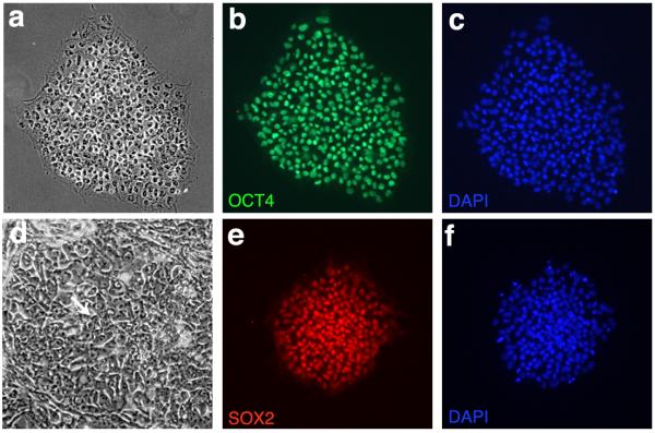
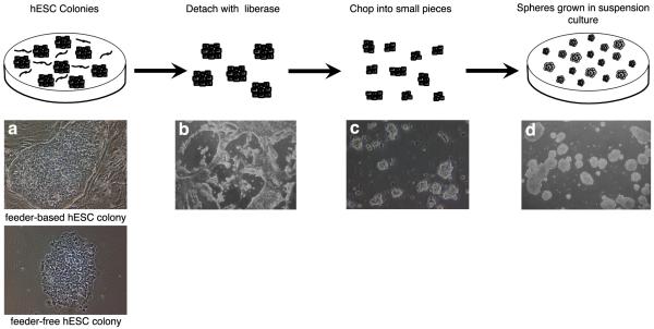
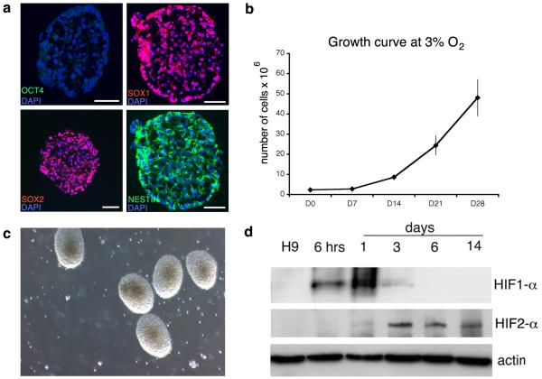
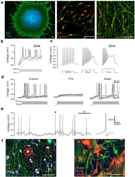
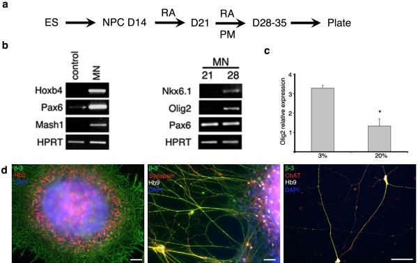
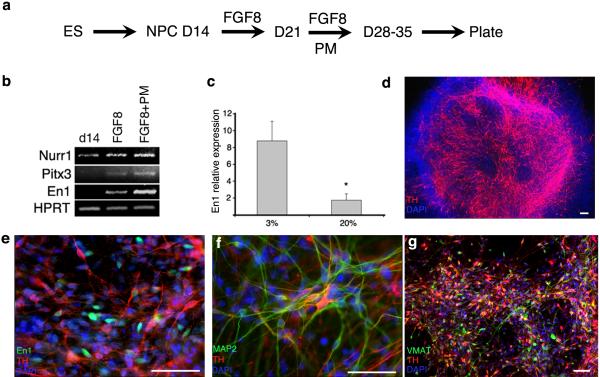
This protocol has been designed to generate neural precursor cells (NPCs) from human embryonic stem cells (hESCs) using a physiological oxygen (O(2)) level of 3% (previously termed hypoxia) and chemically defined conditions. The first stage involves suspension culture of hESC colonies at 3% O(2), where they acquire a neuroepithelial identity over a period of 2 weeks. This timescale is comparable to that observed at 20% O(2), but survival is enhanced. Sequential application of retinoic acid and purmorphamine (PM), from day 14 to day 28, directs differentiation toward spinal motor neurons. Alternatively, addition of fibroblast growth factor-8 and PM generates midbrain dopaminergic neurons. OLIG2 (encoding oligodendrocyte lineage transcription factor 2) induction in motor neuron precursors is twofold greater than that at 20% O(2), whereas EN1 (encoding engrailed homeobox 1) expression is enhanced fivefold. NPCs (at 3% O(2)) can be differentiated into all three neural lineages, and such cultures can be maintained long term in the absence of neurotrophins. The ability to generate defined cell types at 3% O(2) should represent a significant advancement for in vitro disease modeling and potentially for cell-based therapies.


Figures

Figure One
Figure One
hESC Culture: Colonies demonstrate a uniform, undifferentiated appearance with well defined borders (a) and the characteristic double nucleoli present within individual cells (arrow, d). hESCs label with the pluripotency markers OCT4 (b, c) and SOX2 (e,f).
Figure Two
Figure Two
Procedure for Neural Conversion: Feeder-free or feeder-based hESC colonies (a) are detached by incubation with liberase for 10-30 mins (b). The colonies are chopped into small pieces at 120 μm distances in two directions (c) which then form spheres when grown in suspension culture (d).
Figure Three
Figure Three
NPC Generation at 3% O2: After 14 days in culture, NPCs are generated, expressing SOX2, SOX1 (98.7±0.5%) and NESTIN (97.4±0.3%) with loss of OCT4 (1.1±0.7%) (a). NPCs grow rapidly at 3% O2 (b) and, by D14, look healthy, with a phase-bright appearance, well-rounded borders and few dead cells in the background (c). HIF-1α expression is only transiently observed on transfer to the low oxygen environment, whilst HIF-2α appears later but expression persists (d). Scale bar = 50 μm. Figure 3d reproduced from Stacpoole et al, 2011, Cell Death and Differentiation.
Figure Four
Figure Four
Functional Differentiation at 3% O2: β-III TUBULIN positive processes emerge from spheres plated at day 30 for 24 hrs. SYNAPSIN can be detected after 48 hrs of terminal differentiation and MAP2 after 5 days (a). Action potentials can be evoked by current injection, as early as 10 days after plating (b). After 30 days of terminal differentiation, increasing frequency of action potential firing can be observed with increasing current injection, as expected for functional neurons (c), and these are reversibly blocked by the sodium channel blocker TTX (30 days post plating, n=16)(d). Spontaneous action potentials can also be detected, after terminal differentiation for 30 days (e). Asterisk denotes the action potential which is magnified on the right. At later time points, O4 positive oligodendrocytes as well as GFAP and VIMENTIN positive astrocytes are also observed (f). Scale bar = 50 μm. Part of figure 4a reproduced from Stacpoole et al, 2011, Cell Death and Differentiation.
Figure Five
Figure Five
Spinal Motor Neurons: Sequential application of 0.1 μM RA for 7 days, followed by RA and 1 μM PM for 7-14 days (a) generates spinal motor neuron precursors expressing HOXB4, PAX6, OLIG2 and NKX6.1 (b). OLIG2 induction is 2-fold more efficient than at 20% O2 (c). HB9 and β-III positive motor neurons are present after 48 hrs of terminal differentiation, and mature to express SYNAPSIN and ChAT (d). Scale bar = 50 μm. Figure 5b reproduced from Stacpoole et al, 2011, Cell Death and Differentiation.
Figure Six
Figure Six
Midbrain Dopaminergic Neurons: Sequential application of 100 ng/ ml FGF-8 for 7 days followed by FGF-8 and 1 μM PM for 7-14 days (a) generates cells which express the midbrain marker EN1 and NURR1 and PITX3, which are required for the development of dopaminergic neurons of the substantia nigra (b). EN1 induction is 5-fold greater than at 20% O2 (c). Large numbers of TH-positive neurons appear after 10days (d), with co-staining of EN1 and TH apparent after 48hrs of terminal differentiation (e). These TH-positive neurons mature to express MAP2 (f) and VMAT (g). Scale bar = 50 μm Figure 6b & c reproduced from Stacpoole et al, 2011, Cell Death and Differentiation.

References

    1. Zhao T, et al. Hypoxia-driven proliferation of embryonic neural stem/progenitor cells--role of hypoxia-inducible transcription factor-1alpha. FEBS J. 2008;275:1824–1834. - PubMed
    1. Erceg S, Ronaghi M, Stojkovic M. Human embryonic stem cell differentiation toward regional specific neural precursors. Stem Cells. 2009;27:78–87. - PMC - PubMed
    1. Hedlund E, Perlmann T. Neuronal cell replacement in Parkinson’s disease. J. Intern. Med. 2009;266:358–371. - PubMed
    1. Conti L, Cattaneo E. Neural stem cell systems: physiological players or in vitro entities? Nat. Rev. Neurosci. 2010;11:176–187. - PubMed
    1. Munoz-Sanjuan I, Brivanlou AH. Neural induction, the default model and embryonic stem cells. Nat. Rev. Neurosci. 2002;3:271–280. - PubMed

Publication types

MeSH terms