

doi: 10.4103/0971-3026.82300.
Emphysematous cholecystitis: Imaging findings in nine patients
Affiliations
- PMID: 21799598
- PMCID: PMC3137852
- DOI: 10.4103/0971-3026.82300
Item in Clipboard
Emphysematous cholecystitis: Imaging findings in nine patients
Indian J Radiol Imaging.
2011 Apr.
Abstract
Emphysematous cholecystitis is a severe form of acute cholecystitis and can be rapidly fatal. We present the imaging features of nine patients with proven emphysematous cholecystitis.
Keywords: CT scan; cholecystitis; emphysematous; gall bladder; radiograph; ultrasound.
Conflict of interest statement
Figures
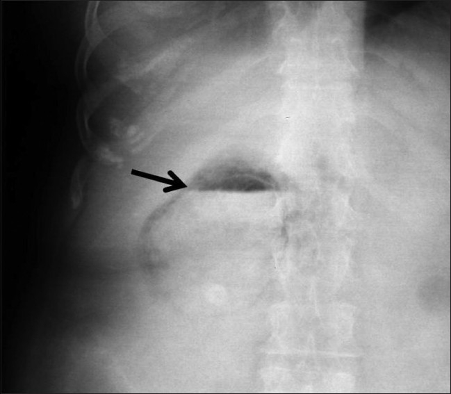
Abdominal radiograph (frontal projection) shows intraluminal air (arrow) with air–fluid levels
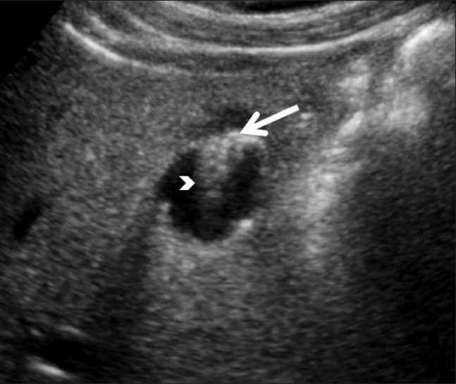
Axial USG image shows a linear echogenic focus (arrow) with a reverberation artifact (arrowhead) at the gall bladder fundus

Sagittal USG image of the gall bladder shows multiple speckled echogenic bands (arrows), with acoustic shadowing within its wall (arrowhead)

Sagittal (A) and axial (B) USG images show sludge (black arrow) and wall thickening (arrow in A) of the gall bladder, consistent with acute cholecystitis with echogenic shadowing foci (arrow in B) within the biliary system

Axial CT scan shows air (arrow) in the gall bladder lumen with an air–fluid level

Axial CT scan shows gall bladder wall thickening (arrow), with intramural air (arrowhead)

Axial CT scan shows intramural air in the gall bladder (arrow), with cholelithiasis (arrowhead)

Axial CT scan shows intrahepatic abscess (arrow), with a focus of air (arrowhead). Also note foci of free intraperitoneal air (curved arrow)

Axial CT scan (A) shows an inflamed gall bladder with questionable air in the gall bladder wall (arrow). Coronal reformation (B) confirms air within the gall bladder wall (arrow) suggesting emphysematous cholecystitis

Adenomyomatosis of the gall bladder: Axial USG image (A) shows an echogenic focus within the gallbladder wall producing a ‘V’-shaped comet tail reverberation artifact (arrow). MRCP image (B) in the same patient shows multiple diverticuli arising from the gall bladder wall (arrow) representing Rokitansky-Aschoff sinuses
References
-
- May RE, Strong R. Acute emphysematous cholecystitis. Br J Surg. 1971;58:453–8. - PubMed
-
- Mentzer RM, Jr, Golden GT, Chandler JG, Horsley JS., 3rd A comparative appraisal of emphysematous cholecystitis. Am J Surg. 1975;129:10–5. - PubMed
-
- Hegner CF. Gaseous pericholecystitis and cholelithiasis. Arch Surg. 1931;22:993–1000.
-
- Mentzer RM, Golden GT, Chandler JG, Horsley JS. A Comparative Appraisal of emphysematous cholecystitis. Am J Surg. 1975;129:10–5. - PubMed
-
- Stolz A. Ubergasbildung in derGollinweger. Arch Pathol Anat. 1901;165:90–123.
LinkOut - more resources
Full Text Sources