Back bugged: A case of sacral hydatid cyst
- PMID: 21799621
- PMCID: PMC3137835
- DOI: 10.4103/0976-3147.63104
Back bugged: A case of sacral hydatid cyst
Abstract
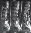
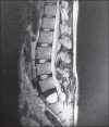
Hydatid cyst of bone constitutes only 0.5 - 2% of all hydatidoses. The thoracic spine is the most common site of spinal hydatidoses. Primary hydatid cyst of the sacral spinal canal is rare. A 23-year-old gentleman had back pain five years ago. At that time he was evaluated and found to have a small cyst in S1 spinal canal, which was presumed to be a benign Tarlov's cyst; and no treatment was offered. He continued to have back pain and also developed sciatica on the right side. Neurological examination presently revealed right S1 radiculopathy. Magnetic resonance imaging (MRI) showed a large multiloculated cystic lesion extending from L5 to S2 spinal canal with bone erosion, both anteriorly and posteriorly. He underwent L5 to S2 laminectomy and excision of multiple cysts. The whole cyst was excised and cavity irrigated with sterilized formalin. A laparoscope was introduced in the cavity to look for extension into the pelvis and to confirm complete excision. Postoperatively, the patient received albendazole for two months. At 16 months follow-up the patient was asymptomatic. Hydatid cyst of sacrum is rare and can be missed at initial presentation. If the patient with a cystic lesion of sacral continues to have symptoms the diagnosis should be revaluated and prompt treatment should be offered.
Keywords: Cestode; echinococcus; hydatid; sacrum; spine.
Conflict of interest statement
Figures


References
-
- Abbassioun K, Amirjamshidi A. Diagnosis and management of hydatid cyst of the central nervous system: Part 2: Hydatid cysts of the skull, orbit, and spine. Neuro Quart. 2001;11:10–6.
-
- Murray RO, Hddad F. Hydatid disease of the spine. J Bone Joint Surg Br. 1959;4:499–506. - PubMed
-
- Sapkas GS, Machinis TG, Chloros GD, Fountas KN, Themistocleous GS, Vrettakos G. Spinal hydatid disease, a rare but existent pathological entity: Case report and review of the literature. South Med J. 2006;99:178–83. - PubMed
-
- Rumana M, Mahadevan A, Nayil Khurshid M, Kovoor JM, Yasha TC, Santosh V, et al. Cestode parasitic infestation: intracranial and spinal hydatid disease: A clinicopathological study of 29 cases from South India. Clin Neuropathol. 2006;25:98–104. - PubMed
Publication types
LinkOut - more resources
Full Text Sources
