

Scaffold fabrication in a perfusion culture microchamber array chip by O(2) plasma bonding of poly(dimethylsiloxane) protected by a physical mask
- PMID: 21799711
- PMCID: PMC3145230
- DOI: 10.1063/1.3576933
Scaffold fabrication in a perfusion culture microchamber array chip by O(2) plasma bonding of poly(dimethylsiloxane) protected by a physical mask
Abstract
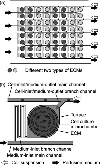
Extracellular matrix (ECM) proteins are required for cell culture. In this paper, we report the use of O(2) plasma bonding to fabricate a perfusion culture microchamber array chip with identical-size ECM spots in the isolated microchambers. The chip was fabricated by assembly of two poly(dimethylsiloxane) (PDMS) layers, a microfluidic network layer, and an ECM array layer, which were aligned and then bonded by O(2) plasma oxidation with protection of the ECM microarray with a physical mask made from PDMS. We successfully cultivated Chinese hamster ovary K1 cells in the microchambers with fibronectin. In the fibronectin microchambers, the cells adhered and extended after 12 h of static culture and then grew over the course of 1 d of perfusion culture.
Figures


Similar articles
-
On-chip cell culture on a microarray of extracellular matrix with surface modification of poly(dimethylsiloxane).Biotechnol J. 2010 May;5(5):463-9. doi: 10.1002/biot.201000021. Biotechnol J. 2010. PMID: 20349452
-
Microfluidic perfusion culture of human induced pluripotent stem cells under fully defined culture conditions.Biotechnol Bioeng. 2014 May;111(5):937-47. doi: 10.1002/bit.25150. Epub 2013 Nov 30. Biotechnol Bioeng. 2014. PMID: 24222619
-
Microfluidic perfusion culture.Methods Mol Biol. 2014;1104:251-63. doi: 10.1007/978-1-62703-733-4_17. Methods Mol Biol. 2014. PMID: 24297421
-
Compartmentalized microfluidic perfusion system to culture human induced pluripotent stem cell aggregates.J Biosci Bioeng. 2017 Aug;124(2):234-241. doi: 10.1016/j.jbiosc.2017.03.014. Epub 2017 Apr 20. J Biosci Bioeng. 2017. PMID: 28434976
-
Poly(dimethylsiloxane) as a material for fabricating microfluidic devices.Acc Chem Res. 2002 Jul;35(7):491-9. doi: 10.1021/ar010110q. Acc Chem Res. 2002. PMID: 12118988 Review.
Cited by
-
Preface to Special Topic: Microfluidics in cell biology and tissue engineering.Biomicrofluidics. 2011 Jun;5(2):22101. doi: 10.1063/1.3594781. Epub 2011 Jun 29. Biomicrofluidics. 2011. PMID: 21799707 Free PMC article.
-
Modulating the foreign body response of implants for diabetes treatment.Adv Drug Deliv Rev. 2021 Jul;174:87-113. doi: 10.1016/j.addr.2021.01.011. Epub 2021 Jan 21. Adv Drug Deliv Rev. 2021. PMID: 33484736 Free PMC article. Review.
References
LinkOut - more resources
Full Text Sources
Research Materials