Automated microinjection of recombinant BCL-X into mouse zygotes enhances embryo development
- PMID: 21799744
- PMCID: PMC3140481
- DOI: 10.1371/journal.pone.0021687
Automated microinjection of recombinant BCL-X into mouse zygotes enhances embryo development
Abstract
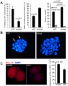
Progression of fertilized mammalian oocytes through cleavage, blastocyst formation and implantation depends on successful implementation of the developmental program, which becomes established during oogenesis. The identification of ooplasmic factors, which are responsible for successful embryo development, is thus crucial in designing possible molecular therapies for infertility intervention. However, systematic evaluation of molecular targets has been hampered by the lack of techniques for efficient delivery of molecules into embryos. We have developed an automated robotic microinjection system for delivering cell impermeable compounds into preimplantation embryos with a high post-injection survival rate. In this paper, we report the performance of the system on microinjection of mouse embryos. Furthermore, using this system we provide the first evidence that recombinant BCL-XL (recBCL-XL) protein is effective in preventing early embryo arrest imposed by suboptimal culture environment. We demonstrate that microinjection of recBCL-XL protein into early-stage embryos repairs mitochondrial bioenergetics, prevents reactive oxygen species (ROS) accumulation, and enhances preimplantation embryo development. This approach may lead to a possible treatment option for patients with repeated in vitro fertilization (IVF) failure due to poor embryo quality.
Conflict of interest statement
Figures


Similar articles
-
A Review of Automated Microinjection of Zebrafish Embryos.Micromachines (Basel). 2018 Dec 24;10(1):7. doi: 10.3390/mi10010007. Micromachines (Basel). 2018. PMID: 30586877 Free PMC article. Review.
-
Osmolarity at early culture stage affects development and expression of apoptosis related genes (Bax-alpha and Bcl-xl) in pre-implantation porcine NT embryos.Mol Reprod Dev. 2008 Mar;75(3):464-71. doi: 10.1002/mrd.20785. Mol Reprod Dev. 2008. PMID: 17948237
-
Limited relationships between reactive oxygen species levels in culture media and zygote and embryo development.J Assist Reprod Genet. 2019 Feb;36(2):325-334. doi: 10.1007/s10815-018-1363-6. Epub 2018 Nov 10. J Assist Reprod Genet. 2019. PMID: 30415468 Free PMC article.
-
Evidence for the requirement of autocrine growth factors for development of mouse preimplantation embryos in vitro.Biol Reprod. 1997 Jan;56(1):229-37. doi: 10.1095/biolreprod56.1.229. Biol Reprod. 1997. PMID: 9002654
-
Comparative analysis of mouse and human preimplantation development following POU5F1 CRISPR/Cas9 targeting reveals interspecies differences.Hum Reprod. 2021 Apr 20;36(5):1242-1252. doi: 10.1093/humrep/deab027. Hum Reprod. 2021. PMID: 33609360
Cited by
-
High fat diet induced developmental defects in the mouse: oocyte meiotic aneuploidy and fetal growth retardation/brain defects.PLoS One. 2012;7(11):e49217. doi: 10.1371/journal.pone.0049217. Epub 2012 Nov 12. PLoS One. 2012. PMID: 23152876 Free PMC article.
-
A Review of Automated Microinjection of Zebrafish Embryos.Micromachines (Basel). 2018 Dec 24;10(1):7. doi: 10.3390/mi10010007. Micromachines (Basel). 2018. PMID: 30586877 Free PMC article. Review.
-
Nanofountain probe electroporation (NFP-E) of single cells.Nano Lett. 2013 Jun 12;13(6):2448-57. doi: 10.1021/nl400423c. Nano Lett. 2013. PMID: 23650871 Free PMC article.
-
Establishment of an integrated automated embryonic manipulation system for producing genetically modified mice.Sci Rep. 2021 Jun 3;11(1):11770. doi: 10.1038/s41598-021-91148-9. Sci Rep. 2021. PMID: 34083640 Free PMC article.
-
High Throughput and Highly Controllable Methods for In Vitro Intracellular Delivery.Small. 2020 Dec;16(51):e2004917. doi: 10.1002/smll.202004917. Epub 2020 Nov 25. Small. 2020. PMID: 33241661 Free PMC article. Review.
References
-
- Ziebe S, Petersen K, Lindenberg S, Andersen AG, Gabrielsen A, et al. Embryo morphology or cleavage stage: how to select the best embryos for transfer after in-vitro fertilization. Hum Reprod. 1997;12:1545–1549. - PubMed
-
- Hardy K, Stark J. Mathematical models of the balance between apoptosis and proliferation. Apoptosis. 2002;7:373–381. - PubMed
-
- Goddard MJ, Pratt HP. Control of events during early cleavage of the mouse embryo: an analysis of the '2-cell block'. J Embryol Exp Morphol. 1983;73:111–133. - PubMed
-
- Maleszewski M, Borsuk E, Koziak K, Maluchnik D, Tarkowski AK. Delayed sperm incorporation into parthenogenetic mouse eggs: sperm nucleus transformation and development of resulting embryos. Mol Reprod Dev. 1999;54:303–310. - PubMed
Publication types
MeSH terms
Substances
Grants and funding
LinkOut - more resources
Full Text Sources
Other Literature Sources
Research Materials

