. 2011;6(7):e22438.
doi: 10.1371/journal.pone.0022438. Epub 2011 Jul 22.

Krüppel-like factor 6 expression changes during trophoblast syncytialization and transactivates ßhCG and PSG placental genes

Affiliations

Krüppel-like factor 6 expression changes during trophoblast syncytialization and transactivates ßhCG and PSG placental genes

Ana C Racca et al. PLoS One. 2011.

Abstract

Background: Krüppel-like factor-6 (KLF6) is a widely expressed member of the Sp1/KLF family of transcriptional regulators involved in differentiation, cell cycle control and proliferation in several cell systems. Even though the highest expression level of KLF6 has been detected in human and mice placenta, its function in trophoblast physiology is still unknown.

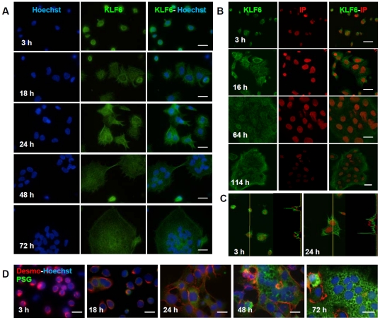
Methodology/principal findings: Herein, we explored KLF6 expression and sub-cellular distribution in human trophoblast cells differentiating into the syncytial pathway, and its role in the regulation of genes associated with placental development and pregnancy maintenance. Confocal immunofluorescence microscopy demonstrated that KLF6 is expressed throughout human cytotrophoblast differentiation showing no evident modifications in its nuclear and cytoplasmic localization pattern. KLF6 transcript and protein peaked early during the syncytialization process as determined by qRT-PCR and western blot assays. Overexpression of KLF6 in trophoblast-derived JEG-3 cells showed a preferential nuclear signal correlating with enhanced expression of human β-chorionic gonadotropin (βhCG) and pregnancy-specific glycoprotein (PSG) genes. Moreover, KLF6 transactivated βhCG5, PSG5 and PSG3 gene promoters. Deletion of KLF6 Zn-finger DNA binding domain or mutation of the consensus KLF6 binding site abolished transactivation of the PSG5 promoter.

Conclusions/significance: Results are consistent with KLF6 playing a role as transcriptional regulator of relevant genes for placental differentiation and physiology such as βhCG and PSG, in agreement with an early and transient increase of KLF6 expression during trophoblast syncytialization.


Conflict of interest statement

Competing Interests: The authors have declared that no competing interests exist.

Figures

Figure 1
Figure 1. KLF6 protein expression throughout trophoblast cell differentiation.
A- Isolated mononuclear villous CTB cells cultured during the indicated hours and stained for KLF6 immunofluorescence detection (middle panels) with the polyclonal R-173 (green) anti-KLF6 antibody. Nuclei were counterstained with Hoechst 33342 dye (blue) and the overlay is shown (right panels). B- Confocal microscopy imaging of KLF6 at the indicated time points of the differentiation process. KLF6 was labelled with the polyclonal R-173 antibody (left panels) and DNA was stained with propidium iodide (IP) (middle panel). Overlay is shown in the right panels. C- Fluorescence intensity profile of KLF6 (green) and IP (red) along the yellow line shown in the confocal microscopy images. D- Morphological and biochemical differentiation of isolated mononuclear CTB cells were confirmed by the disappearance of desmoplakin intercellular staining (red), the appearance of multinucleated structures and the expression of PSG proteins (green). Original magnification, x1000. Scale bar, 10 µm. Immunofluorescence assays were performed with at least three different CTB purifications and representative figures are shown.
Figure 2
Figure 2. KLF6 transcript and protein levels increase during trophoblast cell differentiation.
A- KLF6 mRNA expression was quantified by qRT-PCR (ABI 7500, Applied Biosystems) in CTB cells isolated from three to eight normal term placentas, and cultured in differentiating medium during the indicated times. Results were normalized to cyclophilin A and expressed according to the 2−ΔΔCt method using as calibrator the expression level at 0 h. Results are depicted as boxplot graphs representing the medians (horizontal bars), the 25–75th percentile interquartile range (box limits), and the lowest and highest values (whiskers) of three to eight experiments performed in triplicates. Inter-group comparisons were made using the Kruskal-Wallis one-way Analysis of variance (ANOVA) with the Dunn's multiple comparisons post-hoc test of statistical significance. *p<0.05 vs 0 h, #p<0.05 vs 2 h. B- Protein extracts (60 µg) prepared from CTB cells cultured for the indicated hours were subjected to western blot analysis using anti-KLF6 and α-tubulin antibodies as described in Materials and Methods . A representative blot is shown. The bar graph represents the densitometric quantification of KLF6/α-tubulin ratio of three independent experiments expressed as mean ±SEM. *p<0.05 vs 0 h, ‡p<0.05 vs 16 h. (Kruskal-Wallis, Dunn's).
Figure 3
Figure 3. KLF6 regulates mRNA and protein expression of placental genes.
A- JEG-3 cells were cultured in the presence of 1 µM methotrexate to induce cell differentiation during the indicted times and KLF6 mRNA expression was quantified by qRT-PCR (ABI 7500, Applied Biosystems). B- JEG-3 cells transfected with the empty (white bars) or the KLF6 expression vector (black bars) were harvested 24 h after transfection and PSG, βhCG and GCM1 gene expression was quantified by qRT- PCR (ABI 7500, Applied Biosystems). For A and B, results were normalized to cyclophilin A and expressed according to the 2−ΔΔCt method using as calibrator the mRNA level obtained from the control condition. Data are presented as mean ±SEM of three independent experiments performed in triplicates and a one-sample t-test was used to determine whether experimental values were significantly different from the control value set as 1 (*p<0.05). C- Western blot detection of KLF6, PSG, βhCG and GCM1 in protein extracts of JEG-3 cells transfected with the empty (left lane) or KLF6 expression vector (right lane). α-tubulin was used as a loading control in each assay. Representative western blots are shown and the bar graph shows the densitometric analysis of three different experiments. (*p<0.05) D- JEG-3 cells were transiently transfected with the KLF6 expression vector and 24 h later they were immunostained for the detection of KLF6 (red) and βhCG (green) with a monoclonal anti-KLF6 and a polyclonal anti-βhCG antibodies, respectively. Nuclei were counterstained with Hoechst 33342 dye (blue), and the merge of the three channels is shown on the right side. Bar = 10 µm. Original magnification: ×1000. Representative images from three independent transfections are shown. Arrowheads, JEG-3 cells overexpressing KLF6; arrows, cells positive for βhCG.
Figure 4
Figure 4. KLF6 transactivates βhCG, PSG5 and PSG3 genes in human trophoblastic cells.
The human wild-type KLF6 (KLF6) expression plasmid, the Zn-finger deleted KLF6 (ΔZF 1–3) construct or the empty (-) expression vector were cotransfected into JEG-3 cells together with the Renilla reporter plasmid and (A) the human βhCG5 promoter luciferase reporter plasmids: −3700βhCG5-luc and −345βhCG5-luc or (B) the PSG5 or PSG3 promoter constructs: PB5-luc, PB3-luc, UB5-luc and UB5mut-luc, as depicted on the left. PSG5 and PSG3 consensus KLF6 binding sites, as well as putative KLF6 binding motifs in the βhCG5 gene are depicted as circles. Luciferase activity was normalized to Renilla activity. The KLF6 expression plasmid quantity used is indicated. Results are expressed as fold activation respect to each promoter construct co-transfected with the empty vector. Data from three or four experiments performed in duplicate are shown (mean ±SD). * Statistically significant difference (p<0.05) between the fold activation of each construct in the presence respect to the absence of the expression vectors is shown.

References

    1. Huppertz B, Borges M. Placenta trophoblast fusion. Methods Mol Biol. 2008;475:135–147. - PubMed
    1. Baczyk D, Drewlo S, Proctor L, Dunk C, Lye S, et al. Glial cell missing-1 transcription factor is required for the differentiation of the human trophoblast. Cell Death Differ. 2009;16:719–727. - PubMed
    1. Yang M, Lei ZM, Rao Ch V. The central role of human chorionic gonadotropin in the formation of human placental syncytium. Endocrinology. 2003;144:1108–1120. - PubMed
    1. Knofler M, Saleh L, Bauer S, Galos B, Rotheneder H, et al. Transcriptional regulation of the human chorionic gonadotropin beta gene during villous trophoblast differentiation. Endocrinology. 2004;145:1685–1694. - PubMed
    1. Cole LA. New discoveries on the biology and detection of human chorionic gonadotropin. Reprod Biol Endocrinol. 2009;7:8. - PMC - PubMed

Publication types

MeSH terms