Comparative Study
. 2011;6(7):e22527.
doi: 10.1371/journal.pone.0022527. Epub 2011 Jul 25.

A genome-wide comparative evolutionary analysis of herpes simplex virus type 1 and varicella zoster virus

Affiliations
Comparative Study

A genome-wide comparative evolutionary analysis of herpes simplex virus type 1 and varicella zoster virus

Peter Norberg et al. PLoS One. 2011.

Abstract

Herpes simplex virus type 1 (HSV-1) and varicella zoster virus (VZV) are closely related viruses causing lifelong infections. They are typically associated with mucocutaneous or skin lesions, but may also cause severe neurological or ophthalmic diseases, possibly due to viral- and/or host-genetic factors. Although these viruses are well characterized, genome-wide evolutionary studies have hitherto only been presented for VZV. Here, we present a genome-wide study on HSV-1. We also compared the evolutionary characteristics of HSV-1 with those for VZV. We demonstrate that, in contrast to VZV for which only a few ancient recombination events have been suggested, all HSV-1 genomes contain mosaic patterns of segments with different evolutionary origins. Thus, recombination seems to occur extremely frequent for HSV-1. We conclude by proposing a timescale for HSV-1 evolution, and by discussing putative underlying mechanisms for why these otherwise biologically similar viruses have such striking evolutionary differences.


Conflict of interest statement

Competing Interests: The authors have declared that no competing interests exist.

Figures

Figure 1
Figure 1. HSV-1 phylogeny.
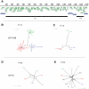
(A) Schematic illustration of the HSV-1 genome. The thick black bar in the bottom depicts the regions that were included in the analysis after all repeat regions and gaps were removed. Nucleotide positions refer to the laboratory strain 17. (B, C, D and E) Phylogenetic networks (splits networks). Networks B and C are based only on US7 and US8, which were the genes analyzed previously by Norberg et al . All strains with recombination crossovers in this region were excluded in order to illustrate the division of the non-recombinant strains into the three distinct evolutionary clades A, B and C. Network B also includes the strains used by Norberg et al, and network C includes only the strains sequenced for this study. Network D is based on the complete genome, including only the strains used in network C (i.e. without recombination crossovers in US7 or US8). Here, recombination crossovers in other parts of the genome prohibit distinct classification of the strains into three distinct clades. Finally, network E is based on the complete genome, including all strains. The network reveals massive recombination and it is irrelevant to attempt to classify a complete genome into any of the three phylogenetic clades A, B or C. The statistical significance for recombination is shown for each dataset.
Figure 2
Figure 2. HSV-1 recombination analysis.
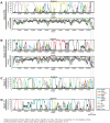
Recombination analysis using Bootscan and SimPlot. A and B illustrate the bootscan and SimPlot analysis of query sequences 2762 and 4-J1037, respectively, which were classified as non-recombinants based on the US7–US8 regions. Bootscan plots demonstrate highly fragmented genomes as a result of recombination. Similarity plots demonstrate the sequence similarity between the query sequence and the other sequences. C and D depicts the bootscan analysis of the reference strains F and 17. Nucleotide positions refer to the sequence alignment excluding gaps and repeat regions.
Figure 3
Figure 3. HSV-1 timescale estimation based on the genes US7 and US8.
Estimated timescale for the HSV-1 evolution. Each major branch point in the phylogenetic tree is marked with a dotted line to the scale-bar below. The 95% confidence intervals for each predicted time since divergence are denoted with purple bars. The increased number of branch points in each clade is highlighted with green background. Posterior probabilities for the major clades are shown.
Figure 4
Figure 4. VZV phylogeny.
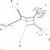
Phylogenetic network (splits network) of available complete genome VZV sequences. The strains are divided into clades 1 to 5, and the deep lineage reticulate topology is caused by a few ancient recombination events described previously. The network also presents a reticulate pattern within clade 1 caused by a putative recent recombination event involving strain SVETA.

References

    1. Roizman B, Sears AE. Herpes simplex viruses and their replication. In: Fields BN, Knipe DM, Howley PM, et al., editors. Virology. Philadelphia: Lippincott-Raven; 1996. pp. 2231–2295.
    1. Lowhagen GB, Tunback P, Bergstrom T. Proportion of herpes simplex virus (HSV) type 1 and type 2 among genital and extragenital HSV isolates. Acta Derm Venereol. 2002;82:118–120. - PubMed
    1. Bergstrom T, Alestig K, Svennerholm B, Horal P, Skoldenberg B, et al. Neurovirulence of herpes simplex virus types 1 and 2 isolates in diseases of the central nervous system. Eur J Clin Microbiol Infect Dis. 1990;9:751–757. - PMC - PubMed
    1. Dix RD, McKendall RR, Baringer JR. Comparative neurovirulence of herpes simplex virus type 1 strains after peripheral or intracerebral inoculation of BALB/c mice. Infect Immun. 1983;40:103–112. - PMC - PubMed
    1. Whitley R, Lakeman AD, Nahmias A, Roizman B. Dna restriction-enzyme analysis of herpes simplex virus isolates obtained from patients with encephalitis. N Engl J Med. 1982;307:1060–1062. - PubMed

Publication types