. 2011;6(7):e22542.
doi: 10.1371/journal.pone.0022542. Epub 2011 Jul 20.

Identification of RNF213 as a susceptibility gene for moyamoya disease and its possible role in vascular development

Affiliations

Identification of RNF213 as a susceptibility gene for moyamoya disease and its possible role in vascular development

Wanyang Liu et al. PLoS One. 2011.

Abstract

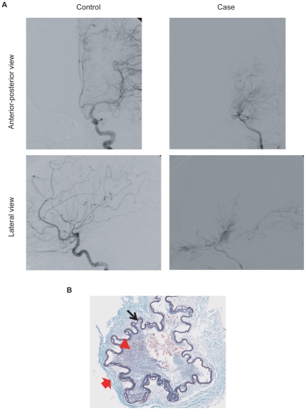
Background: Moyamoya disease is an idiopathic vascular disorder of intracranial arteries. Its susceptibility locus has been mapped to 17q25.3 in Japanese families, but the susceptibility gene is unknown.

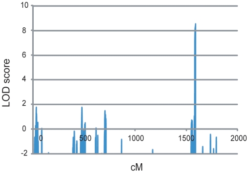
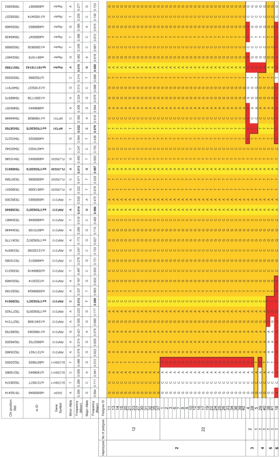
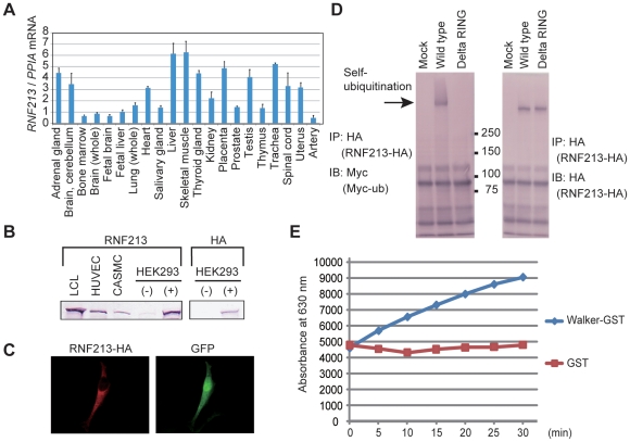
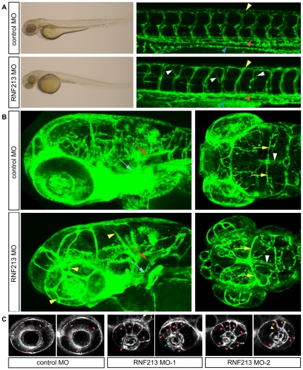
Methodology/principal findings: Genome-wide linkage analysis in eight three-generation families with moyamoya disease revealed linkage to 17q25.3 (P<10(-4)). Fine mapping demonstrated a 1.5-Mb disease locus bounded by D17S1806 and rs2280147. We conducted exome analysis of the eight index cases in these families, with results filtered through Ng criteria. There was a variant of p.N321S in PCMTD1 and p.R4810K in RNF213 in the 1.5-Mb locus of the eight index cases. The p.N321S variant in PCMTD1 could not be confirmed by the Sanger method. Sequencing RNF213 in 42 index cases confirmed p.R4810K and revealed it to be the only unregistered variant. Genotyping 39 SNPs around RNF213 revealed a founder haplotype transmitted in 42 families. Sequencing the 260-kb region covering the founder haplotype in one index case did not show any coding variants except p.R4810K. A case-control study demonstrated strong association of p.R4810K with moyamoya disease in East Asian populations (251 cases and 707 controls) with an odds ratio of 111.8 (P = 10(-119)). Sequencing of RNF213 in East Asian cases revealed additional novel variants: p.D4863N, p.E4950D, p.A5021V, p.D5160E, and p.E5176G. Among Caucasian cases, variants p.N3962D, p.D4013N, p.R4062Q and p.P4608S were identified. RNF213 encodes a 591-kDa cytosolic protein that possesses two functional domains: a Walker motif and a RING finger domain. These exhibit ATPase and ubiquitin ligase activities. Although the mutant alleles (p.R4810K or p.D4013N in the RING domain) did not affect transcription levels or ubiquitination activity, knockdown of RNF213 in zebrafish caused irregular wall formation in trunk arteries and abnormal sprouting vessels.

Conclusions/significance: We provide evidence suggesting, for the first time, the involvement of RNF213 in genetic susceptibility to moyamoya disease.


Conflict of interest statement

Competing Interests: Kyoto University has applied to the Patent Office, Japan, for a patent on this gene. The authors confirm that this does not alter their adherence to all the PLos One policies on sharing data and materials.

Figures

Figure 1
Figure 1. Clinical features of moyamoya disease.
(A) An anterior-posterior and lateral views of left internal carotid angiography in a 9-year-old child with moyamoya disease (right) and an 8-year-old control child (left). An occlusion with moyamoya vessels can be seen around the terminal portion of the internal carotid artery and proximal portions of the anterior cerebral artery and middle cerebral artery in the affected child. (B) Intimal hyperplasia in the middle cerebral artery from an autopsy of a 69-year-old case. Intimal hyperplasia (red arrowhead), atrophic media (red arrow) and winding internal elastic lamina (black arrow) can be seen with Elastica Masson staining, ×200.
Figure 2
Figure 2. The eight three-generation families and haplotype construction with fine markers.
For familial index cases in the eight pedigrees, the haplotypes are indicated by the boxes constructed in GENEHUNTER . The smallest disease haplotype spanning from D17S1806 to rs2280147 was estimated by the region common to both 1411 in the pedigree 2 and 132 in pedigree 20.
Figure 3
Figure 3. Genome-wide linage analysis uniquely identified a locus on 17q25.3 for the eight families.
Genome wide linkage analysis showed a highest LOD score (8.46) at D17S784 in the locus 17q25.3.
Figure 4
Figure 4. Transmission patterns of the haplotypes in 42 families.
The numbers in the parentheses indicate the haplotypes transmitted in each family. Haplotype 1 symbolizes non-risk haplotypes collectively. Therefore it comprises various haplotypes. Haplotypes can be seen in Figure 5. Several family members that were genotyped were omitted in the pedigree chart for clarity. Those members and their haplotypes were; two siblings (1,1) for pedigree 7; father (1,1) for pedigree 21; father (1,1) for pedigree 22; father (1, 1) and a sibling (1,1) for pedigree 23; father (1,1), mother (2,1) and a sibling (1,1) for pedigree 26; three siblings (1,1), (1,1) and (1,1) for pedigree 27; father (1,1), three siblings (1,1) (1,1) and (1,1) for pedigree 28; father (1,1), mother (2,1) and a child of 2 (1,1) for pedigree 32; three siblings (1,1), (1,1), and (1,1) for pedigree 33; mother (1,1) for pedigree 34; father (1,1) and mother (2,1) for pedigree 36; a sibling (1,1) for pedigree 39; three siblings (1,1), (4,1) and (1,1) for pedigree 40; father (2,1) for pedigree 41; mother (1,1) for pedigree 42.
Figure 5
Figure 5. Risk haplotypes transmitted in 42 families.
The orange regions represent the haplotype of the index case of pedigree 11. The yellow regions indicate rare variants. The red and white regions represent flanking SNPs and SNPs outside of the founder haplotype, respectively. The minimum founder haplotype fell in a a region in a span of 130 kb covering RNF213 and FLJ35220. The physical positions were referred from Build 37.1.
Figure 6
Figure 6. Genomic structure, domains of RNF213 and variants.
Genomic structure was based on DDBJ/EMBL-Bank/GenBank accession number AB537889. Domain structure was obtained by GeneCards.
Figure 7
Figure 7. Two isoforms of alternative splicing variants of RNF213.
We have tested whether exon 4 is read through or not in cDNA isolated from various human tissues and HUVECs. Representative results of human tissue RNAs and HUVEC are shown. A short isoform, which skips the exon 4, has an expected size of 270 bp (AB537889) and a long, which reads exon 4, has an expected size of 417 bp (NM_020914.4).
Figure 8
Figure 8. Characterization of the RNF213 gene and protein.
(A) RNF213 mRNA expression. Total RNA from the indicated human tissues was reverse-transcribed to cDNA, and real-time quantitative PCR was performed. (B) RNF213 protein expression. LCL, HUVEC, and CASMC were lysed and immunoblotted using an anti-RNF213 antibody. HEK293 cells transiently expressing RNF213-HA (+) or control cells (−) were immunoblotted using anti-RNF213 and anti-HA antibodies. (C) Subcellular localization of RNF213. HeLa cells transiently expressing RNF213-HA were stained with an anti-HA antibody. (D) Self-ubiquitination of RNF213. HEK293 cells transiently expressing RNF213-HA and Myc-ubiquitin (Myc-ub) were lysed and subjected to immunoprecipitation (IP) using an anti-HA antibody, followed by immunoblotting (IB) using an anti-Myc antibody. As a control, immunoblotting was also performed with an anti-HA antibody. (E) ATPase activity of RNF213. Free phosphate released from ATP by the ATPase activity of a recombinant RNF213 fragment (a.a. 2359–2613) tagged with GST was measured using the Malachite Green method. a.a., amino acid. IP, immunoprecipitation. IB, immunoblot.
Figure 9
Figure 9. Depletion of RNF213 causes abnormal vessel sprouting in zebrafish.
Tg(fli-EGFP)y1 embryos at 72 h post-fertilization. (A) Brightfield image of whole embryos (left) and confocal images of tail vessels (right) of embryos injected with a control (scrambled) or RNF213 morpholino (MO). Lateral views, dorsal uppermost, anterior to the left. In RNF213 morphants, abnormal intersegmental vessel sprouting was observed (white arrowheads). Trunk vessels, including the dorsal aorta (red arrowheads), posterior cardinal vein (blue arrowheads) and dorsal longitudinal anastomotic vessels (yellow arrowheads) developed almost normally. (B) Confocal images of intracranial vessels. (Left) Lateral views 20 degrees toward the top; dorsal uppermost, anterior to the left. (Right) Dorsal views, head to the left. The trunk artery, including the basilar artery (red arrows), lateral dorsal aorta (blue arrows), mesencephalic artery (yellow arrows) and dorsal longitudinal vein (white arrowheads) developed almost normally in controls and RNF213 morphants. RNF213 morphants showed abnormal sprouting vessels (yellow arrowheads) and irregular vessel diameter (white arrowheads). (C) Cross-sectional view at the middle of the lens level. In a control morphant, the three branches of the nasal ciliary artery (red arrowheads) drain into the inner optic circle (IOC; blue arrowheads). In two different RNF213 morphants, multiple aberrant vessels drain into the IOC (red arrowheads), and occasionally part of the IOC was missing (yellow arrowhead).

Similar articles

Cited by

References

    1. Takeuchi K, Shimizu K. Hypogenesis of bilateral internal carotid arteries. Brain Nerve. 1957;9:37–43.
    1. Suzuki J, Takaku A. Cerebrovascular “moyamoya” disease. Disease showing abnormal net-like vessels in base of brain. Arch Neurol. 1969;20:288–299. - PubMed
    1. Goto Y, Yonekawa Y. Worldwide distribution of moyamoya disease. Neurol Med Chir (Tokyo) 1992;32:883–886. - PubMed
    1. Kuroda S, Houkin K. Moyamoya disease: current concepts and future perspectives. Lancet Neurol. 2008;7:1056–1066. - PubMed
    1. Miao W, Zhao PL, Zhang YS, Liu HY, Chang Y, et al. Epidemiological and clinical features of Moyamoya disease in Nanjing, China. Clin Neurol Neurosurg. 2010;112:199–203. - PubMed

Publication types

MeSH terms