. 2011;6(7):e22585.
doi: 10.1371/journal.pone.0022585. Epub 2011 Jul 22.

Epigenetic silencing of nucleolar rRNA genes in Alzheimer's disease

Affiliations

Epigenetic silencing of nucleolar rRNA genes in Alzheimer's disease

Maciej Pietrzak et al. PLoS One. 2011.

Abstract

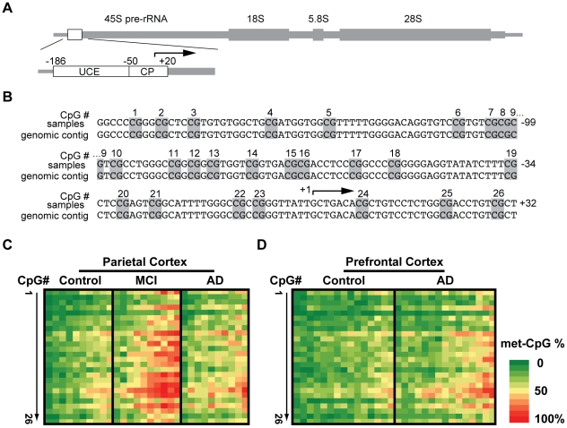
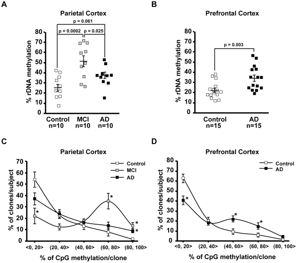
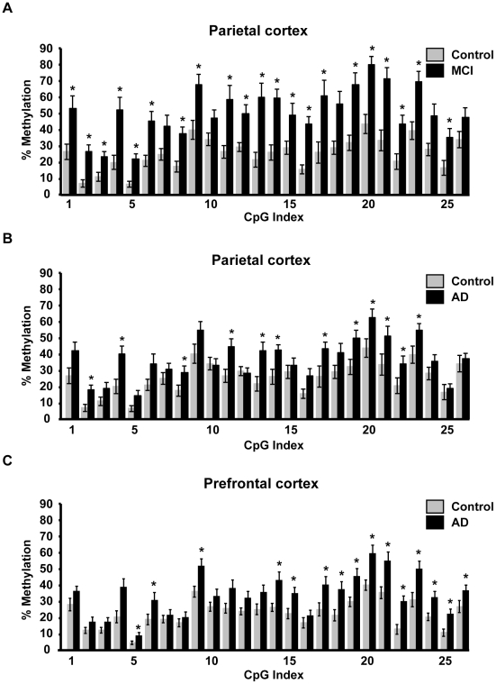
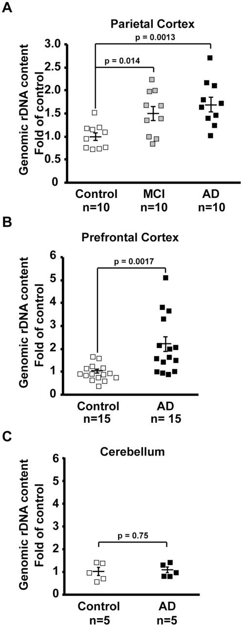
Background: Ribosomal deficits are documented in mild cognitive impairment (MCI), which often represents an early stage Alzheimer's disease (AD), as well as in advanced AD. The nucleolar rRNA genes (rDNA), transcription of which is critical for ribosomal biogenesis, are regulated by epigenetic silencing including promoter CpG methylation.

Methodology/principal findings: To assess whether CpG methylation of the rDNA promoter was dysregulated across the AD spectrum, we analyzed brain samples from 10 MCI-, 23 AD-, and, 24 age-matched control individuals using bisulfite mapping. The rDNA promoter became hypermethylated in cerebro-cortical samples from MCI and AD groups. In parietal cortex, the rDNA promoter was hypermethylated more in MCI than in advanced AD. The cytosine methylation of total genomic DNA was similar in AD, MCI, and control samples. Consistent with a notion that hypermethylation-mediated silencing of the nucleolar chromatin stabilizes rDNA loci, preventing their senescence-associated loss, genomic rDNA content was elevated in cerebrocortical samples from MCI and AD groups.

Conclusions/significance: In conclusion, rDNA hypermethylation could be a new epigenetic marker of AD. Moreover, silencing of nucleolar chromatin may occur during early stages of AD pathology and play a role in AD-related ribosomal deficits and, ultimately, dementia.


Conflict of interest statement

Competing Interests: The authors have declared that no competing interests exist.

Figures

Figure 1
Figure 1. Methyl-cytosine (mC) mapping of the rDNA promoter region.
A, The rDNA gene unit. The location of the promoter region that was selected for bisulfite mapping of methyl cytosines is indicated by a box. That analyzed region included the Upstream Control Element (UCE) and the Core Promoter (CP) which contain 26 CpG methylation sites that are critical for epigenetic regulation of nucleolar transcription (for more detail, see Fig. S1). B. Nucleotide sequence of the human rDNA promoter. The numbers on the right indicate positions relative to the transcription start site at position +1 (arrow); Potentially methylated CpG dinucleotides are marked grey and numbered. C, Bisulfite sequencing analysis of parietal cortex from control-, MCI-, and AD individuals (n = 10 for each group). D. Bisulfite sequencing analysis of prefrontal cortex from control-, AD individuals (n = 15 for each group). In C–D, each column represents one individual. Rows represent each of the potentially methylated 26 CpG sites in the rDNA promoter in 5′ to 3′ orientation. The data (% methylation for a given CpG) are from analyzing 20 unique clones for each subject.
Figure 2
Figure 2. Effects of MCI and AD on mC content in the rDNA promoter.
A–B, Average % mC in the rDNA promoter (% rDNA methylation) in the parietal- (A), and the prefrontal cortex (B). Individual values are depicted by squares; mean values are indicated by the lines intersecting the error bars (SEM); p values and number of analyzed cases (n) are indicated. In the AD-affected prefrontal cortex, the rDNA promoter is hypermethylated. C–D, The distribution of differentially methylated rDNA promoter clones is affected by MCI and AD. For each sample, % rDNA clones were determined within 5 ranges of mC content; a range definition “(20,40>” means more than 20 and less than-, or, equal to 40% mC. Mean values ±SEM are presented for each group of subjects. The bias towards hypomethylated clones is present in controls (ANOVA, p<0.001). That trend is disrupted or flattened in MCI or AD, respectively (two-way ANOVA, interaction between mC range and diagnosis, p<0.001); *, p<0.05 for post-hoc comparisons against in-range controls.
Figure 3
Figure 3. Effects of MCI and AD on distribution of mCpGs across the rDNA promoter.
Percent CpG methylation at each of 26 individual CpG sites located in the rDNA promoter in samples from parietal- (A–B) and prefrontal cortex (C). For the purpose of clarity, parietal cortex data for MCI and AD groups are presented on separate graphs. The controls are the same in both cases. Data are means ± SEM for each analyzed group of cases (n as in Fig. 1); *, p<0.05 (SAM statistics as compared to controls).
Figure 4
Figure 4. Local regression analysis of mC content across the rDNA promoter.
After logarithmic transformation of the data from Fig. 3, the mean differences in methylation of each of 26 individual CpG sites in the rDNA promoter were determined as indicated. A, Local regression analysis of the mean difference between MCI- and control groups (parietal cortex) indicates significant hypermethylation of all CpGs of the rDNA promoter. The circles indicate the actual mean difference values; the black solid line represents a smoothed regression fit of those values across the rDNA promoter region; the green lines indicate the border of the 95% confidence range for the actual values (dashed line) or the smoothed regression (solid line). Note that the black smoothed regression line does not leave the 95% confidence range, indicating a significant hypermethylation trend for all the CpG sites. B–C, Similar analysis of the data for the AD samples from the parietal (B) and the prefrontal cortex (C) identifies AD-associated hypermethylation of large portions of the rDNA promoter.
Figure 5
Figure 5. Increased genomic rDNA content in cerebro-cortical samples from MCI- and AD individuals.
Genomic rDNA was quantified by qRT-PCR; for normalization, tRNA-K-CTT gene was selected as its unclustered copies are scattered throughout several human chromosomes providing a good control of the non-rDNA genome stability. The relative rDNA content was calculated as a ratio of rDNA to tRNA-K-CTT. A, In parietal cortex increased rDNA content was observed both in MCI and AD. B–C, AD- associated increase in rDNA content in the prefrontal cortex (B) but not the cerebellum (C). Individual values are depicted by squares; mean values are indicated by the lines intersecting the error bars (SEM); p values and numbers of analyzed cases (n) are shown.

References

    1. Szyf M. Epigenetics, DNA methylation, and chromatin modifying drugs. Annu Rev Pharmacol Toxicol. 2009;49:243–263. - PubMed
    1. Suzuki MM, Bird A. DNA methylation landscapes: provocative insights from epigenomics. Nat Rev Genet. 2008;9:465–476. - PubMed
    1. Weaver IC, Cervoni N, Champagne FA, D'Alessio AC, Sharma S, et al. Epigenetic programming by maternal behavior. Nat Neurosci. 2004;7:847–854. - PubMed
    1. Chouliaras L, Rutten BP, Kenis G, Peerbooms O, Visser PJ, et al. Epigenetic regulation in the pathophysiology of Alzheimer's disease. Prog Neurobiol. 2010;90:498–510. - PubMed
    1. Siegmund KD, Connor CM, Campan M, Long TI, Weisenberger DJ, et al. DNA methylation in the human cerebral cortex is dynamically regulated throughout the life span and involves differentiated neurons. PLoS One. 2007;2:e895. - PMC - PubMed

Publication types