. 2011 Nov 1;519(16):3346-66.
doi: 10.1002/cne.22730.

Expression of vesicular glutamate transporters type 1 and 2 in sensory and autonomic neurons innervating the mouse colorectum

Affiliations

Expression of vesicular glutamate transporters type 1 and 2 in sensory and autonomic neurons innervating the mouse colorectum

Pablo R Brumovsky et al. J Comp Neurol. .

Abstract

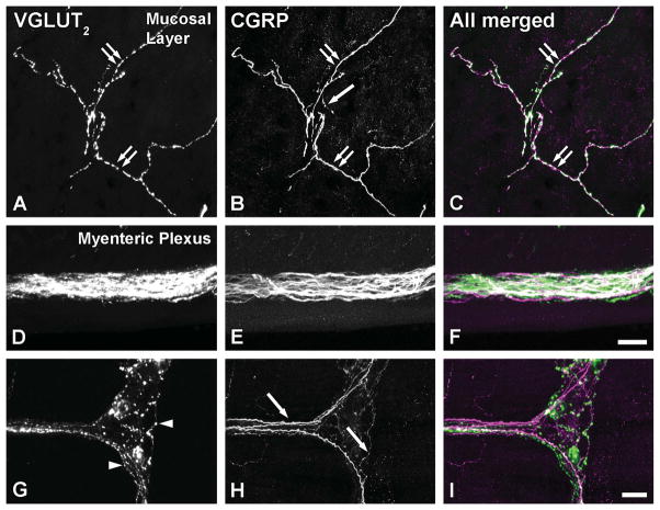
Vesicular glutamate transporters (VGLUTs) have been extensively studied in various neuronal systems, but their expression in visceral sensory and autonomic neurons remains to be analyzed in detail. Here we studied VGLUTs type 1 and 2 (VGLUT(1) and VGLUT(2) , respectively) in neurons innervating the mouse colorectum. Lumbosacral and thoracolumbar dorsal root ganglion (DRG), lumbar sympathetic chain (LSC), and major pelvic ganglion (MPG) neurons innervating the colorectum of BALB/C mice were retrogradely traced with Fast Blue, dissected, and processed for immunohistochemistry. Tissue from additional naïve mice was included. Previously characterized antibodies against VGLUT(1) , VGLUT(2) , and calcitonin gene-related peptide (CGRP) were used. Riboprobe in situ hybridization, using probes against VGLUT(1) and VGLUT(2) , was also performed. Most colorectal DRG neurons expressed VGLUT(2) and often colocalized with CGRP. A smaller percentage of neurons expressed VGLUT(1) . VGLUT(2) -immunoreactive (IR) neurons in the MPG were rare. Abundant VGLUT(2) -IR nerves were detected in all layers of the colorectum; VGLUT(1) -IR nerves were sparse. A subpopulation of myenteric plexus neurons expressed VGLUT2 protein and mRNA, but VGLUT1 mRNA was undetectable. In conclusion, we show 1) that most colorectal DRG neurons express VGLUT(2) , and to a lesser extent, VGLUT(1) ; 2) abundance of VGLUT2-IR fibers innervating colorectum; and 3) a subpopulation of myenteric plexus neurons expressing VGLUT(2). Altogether, our data suggests a role for VGLUT(2) in colorectal glutamatergic neurotransmission, potentially influencing colorectal sensitivity and motility.


Figures

Figure 1
Figure 1
Brightfield optical photomicrographs of lumbar DRG sections, incubated with antiserum to VGLUT1 (A) or VGLUT2 (B), or after coincubation with antibodies and antisense riboprobes against VGLUT1 (C) or VGLUT2 (D) protein and mRNA, respectively. A, B: A number of VGLUT1-IR, large-sized NPs (arrowheads in A), and many VGLUT2-IR NPs of different sizes (arrowheads in B) are detected in DRG sections. NPs lacking any of the two VGLUTs can be seen (asterisks). C, D: Combined riboprobe in situ hybridization–immunohistochemistry experiments reveal that both mRNA and protein for VGLUT1 or VGLUT2 are colocalized in the same DRG NPs (double black arrowheads in C, D, respectively). Neurons lacking both types of signal are also present (asterisks). Scale bar = 50 μm. [Color figure can be viewed in the online issue, which is available at wileyonlinelibrary.com.]
Figure 2
Figure 2
Optical immunofluorescence photomicrographs of sections of T12 (A–C), or S1 (D–I) DRGs incubated with antiserum to VGLUT1 (A) or VGLUT2 (D, G). Retrogradely labeled colorectal DRG neurons containing FB (B, E, H) are shown in red in the merged micrographs (C, F, I). A–C: Only a few colorectal VGLUT1-IR NPs are evident (double arrowheads), but a relatively larger number of noncolorectal VGLUT1-IR, large-sized NPs are present in the section (arrowheads in A) as are colorectal DRG NPs lacking the transporter (arrows in B). D–I: Abundant noncolorectal VGLUT2-IR NPs are present in S1 DRGs, spanning various neuronal sizes (arrowheads in D, G). Virtually all colorectal NPs exhibit VGLUT2-LI (double arrowheads); occasional colorectal NPs apparently lack this transporter (arrow in H). Scale bars = 50 μm in F (applies to A–E); I (applies to G, H). [Color figure can be viewed in the online issue, which is available at wileyonlinelibrary.com.]
Figure 3
Figure 3
Proportions of VGLUT2-IR colorectal NPs lacking (unfilled segments) or coexpressing (filled segments) CGRP in thoracolumbar and lumbosacral DRGs.
Figure 4
Figure 4
Optical immunofluorescence photomicrographs of sections of L6 (A–D) or T12 (E–H) DRGs after coincubation with VGLUT2 (A) or VGLUT1 (E) antiserum and CGRP antiserum (C, G). Retrogradely labeled colorectal neurons containing FB (B, F) are shown in red in the merged micrographs (D, H). A–D: Virtually all VGLUT2-IR colorectal NPs coexpress CGRP (double arrows). Occasional VGLUT2-only colorectal NPs are also evident (white double arrowheads). Additional VGLUT2/CGRP-IR noncolorectal NPs are present (arrows). E–H: Few VGLUT1-IR colonic NPs are present, and these lack CGRP (white double arrowheads). However, most colorectal NPs show CGRP-LI (black double arrowheads). Scale bar = 50 μm. [Color figure can be viewed in the online issue, which is available at wileyonlinelibrary.com.]
Figure 5
Figure 5
Confocal (A, D, G, J, M, P) and optical (B, E, H, K, N, Q) immunofluorescence photomicrographs of sections of the LSC (A–F) or MPG (G–R) after incubation with VGLUT1 (A, G) or VGLUT2 (D, J, M, P) antiserum. Retrogradely labeled colorectal neurons containing FB (B, E, H, K, N, Q) are shown in red in the merged micrographs (C, F, I, L, O, R). A–F: A number of colorectal NPs are detected in the LSC (arrows in B, E). Incubation with the VGLUT1 antiserum failed to reveal any immunostaining, either in neurons or any other ganglionic structure (A, C). In contrast, VGLUT2-LI is detected in fibers (black double arrowhead in D) and what appear to be varicosities (arrowheads in D) in the vicinity of colorectal LSC NPs. However, the latter were always VGLUT2-negative (arrows in E). G–I: Only rare VGLUT1-IR puncta are found in the MPG (arrowheads in G). Note many FB labeled neurons (arrows in H). J–L: In contrast, presence of VGLUT2 in fibers (black double arrowheads), perineuronal baskets (white double arrowheads) (arrows in N), and rare colorectal NPs (double arrows; also shown at higher magnification in the inset in L) was detected in the MPG. M–O: A colonic MPG neuron is contacted by a VGLUT2-IR perineuronal basket, where puncta are easily seen (arrowheads in M). P–R: In addition to a number of fibers (black double arrowheads in P) and puncta (arrowheads in P) seen in the MPG, note the presence of perineuronal VGLUT2-IR baskets around non-colorectal (black arrowheads in P–R) MPG NPs (shown at higher magnification in the inset in R). Scale bars = 50 μm in L (applies to A–K), R (applies to P, Q); 20 μm in insets (L, R); 10 μm in O (applies to M, N). [Color figure can be viewed in the online issue, which is available at wileyonlinelibrary.com.]
Figure 6
Figure 6
Confocal immunofluorescence photomicrographs of transverse sections of the colorectum after incubation with VGLUT1 (A–C) or VGLUT2 (D–F) antiserum (asterisks show the location of the colorectal lumen). A–C: Rare VGLUT1-IR fibers are shown in submucosal (black double arrowheads in A, B) and mucosal (arrow in C) layers in the colorectal wall (inset in B shows fibers at higher magnification). D–F: An abundant VGLUT2-IR neuropil is observed throughout the wall of the colorectum, including the myenteric plexus area (box 1; shown at higher magnification in E) as well as in the mucosa (box 2; shown at higher magnification in F). Note the presence of VGLUT2 in fibers reaching very near the most luminal colorectal epithelium (arrows in D, F). VGLUT2-IR fibers are also found in the submucosal layers (black double arrowhead in D), and create dense neuropil arrangements in the myenteric plexus (double arrows in D, E). Some sparsely located cells show fluorescence due to unspecific binding of the secondary antibody (white arrowheads in D, F). Scale bars = 100 μm in D; 50 μm in B; 20 μm in A, C, E, inset in B; 10 μm in F.
Figure 7
Figure 7
Confocal immunofluorescence photomicrographs of whole-mount colorectal preparations after 24 hours (A–C) or 48h (D–F) free-floating incubation with VGLUT1 (A) or VGLUT2 (B–F) antisera (counterstained in F with propidium iodide, PI) (asterisks show the location of myenteric plexus NPs lacking VGLUT2-positive neuronal cell bodies). A: VGLUT1-LI is absent in the myenteric plexus and surrounding longitudinal muscle. B, C: In contrast, an abundant VGLUT2-IR neuropil is observed in myenteric plexus ganglia, often connected by thick VGLUT2-IR nerve bundles (arrows in B). A high-power magnification of the box in B shows the delicate distribution of VGLUT2, adopting the appearance of varicose endings (arrowheads in C) around VGLUT2-negative myenteric plexus neurons (asterisks). D–F: Forty-eight-hour incubation with the VGLUT2 antibody highlights the presence of a discrete number of myenteric plexus neurons expressing the transporter (black double arrowheads). PI staining confirms the association of this staining pattern with a cellular structure, in this case myenteric plexus neurons. Scale bars = 50 μm in B (applies to A, D); 20 μm in C; 10 μm in E, F. [Color figure can be viewed in the online issue, which is available at wileyonlinelibrary.com.]
Figure 8
Figure 8
Dark- (A, H, C, J) and brightfield (B, I, D, K, E, F, G, L, M) optical photomicrographs of colorectal (A–D, G; H–K) or DRG (E, F, L, M) sections, after hybridization with antisense (A–E; H–L) and sense (F, G, M) riboprobes against VGLUT2 (A–G) or VGLUT1 (H–M) mRNAs. A–D: In transverse sections of the colorectum, VGLUT2 binding is observed only at the level of the myenteric plexus, present in a discrete number of neurons, as seen in a magnified view of the box in A (double arrowheads in B). Myenteric plexus (arrows in A, C) as well as individual neurons (arrowheads in B) lacking VGLUT2 are also apparent. Tangential sections of the myenteric plexus confirm the presence of VGLUT2 in some neurons throughout the plexus (double arrowheads in C, D). Many of the neurons present in each myenteric ganglia lack VGLUT2 (arrowheads in D). E–G: The VGLUT2 antisense riboprobe is effective in showing DRG neuronal expression (double arrowheads in E). In contrast, incubation of the sections with sense riboprobe revealed no VGLUT2 binding in DRG (F) or myenteric plexus (G) sections. H–M: VGLUT1 (H–K) mRNA was virtually absent from all layers of the colorectum. Neurons lacking VGLUT1 were repeatedly seen in the myenteric plexus (arrowheads in I, K). Despite the lack of hybridization in the colon, the VGLUT1 antisense riboprobe was efficient in labeling a number of NPs in DRGs (double arrowheads in L). VGLUT1 binding was absent in sections incubated with the sense riboprobe (M). Scale bars = 100 μm in J (applies to A, H, C); 50 μm in K (applies to B, I, D), in G, M (applies to E, L, F).
Figure 9
Figure 9
Confocal immunofluorescence photomicrographs of parasagittal (A–C; G, H) and transverse (D–F) sections of the colorectum after coincubation with VGLUT2 (A, D, G) and CGRP (B, E, H) antisera (merged figures are shown in C, F, I). A–C: Virtually complete colocalization between VGLUT2 and CGRP is observed in the mucosal layers of the colorectum (double arrows). Additional CGRP-only fibers are also present (arrow in B). D–F: In the myenteric plexus, a dense VGLUT2 and CGRP-IR neuropil is detected. G–I: However, more detailed examination of the populations of VGLUT2-IR (arrowheads in G) and CGRP-IR (arrows in H) fibers shows that they are virtually never overlapping. Scale bars = 20 μm in I (applies to A–C, G, H); in F (applies to D, E). [Color figure can be viewed in the online issue, which is available at wileyonlinelibrary.com.]

References

    1. Abercrombie M. Estimation of nuclear population from microtome sections. Anat Rec. 1946;94:239–247. - PubMed
    1. Adams JC. Biotin amplification of biotin and horseradish peroxidase signals in histochemical stains. J Histochem Cytochem. 1992;40:1457–1463. - PubMed
    1. Aihara Y, Mashima H, Onda H, Hisano S, Kasuya H, Hori T, Yamada S, Tomura H, Yamada Y, Inoue I, Kojima I, Takeda J. Molecular cloning of a novel brain-type Na(+)-dependent inorganic phosphate cotransporter. J Neurochem. 2000;74:2622–2625. - PubMed
    1. Alvarez FJ, Villalba RM, Zerda R, Schneider SP. Vesicular glutamate transporters in the spinal cord, with special reference to sensory primary afferent synapses. J Comp Neurol. 2004;472:257–280. - PubMed
    1. Anlauf E, Derouiche A. Astrocytic exocytosis vesicles and glutamate: a high-resolution immunofluorescence study. Glia. 2005;49:96–106. - PubMed

Publication types

Substances